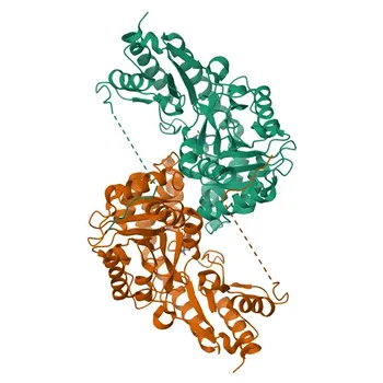
HLA-only antibody screening leaves a dangerous blind spot in transplant risk assessment. Non-HLA antigens such as MICA, AT1R, and vimentin can independently trigger antibody-mediated rejection (AMR) and chronic graft loss—reactions that standard anti-HLA assays completely miss. Integrating high-purity recombinant non-HLA targets into diagnostic panels is critical to capture these immunogenic threats, enabling a truly comprehensive evaluation of organ rejection risk before and after transplantation.
Comprehensive transplant antibody screening must move beyond HLA. Non-HLA targets like MICA, AT1R, and vimentin mediate rejection pathways that standard assays overlook, especially in patients with no detectable donor-specific HLA antibodies. Including these targets gives laboratories the power to identify hidden graft damage, guide early intervention, and improve long-term allograft survival.
The Limitations of an HLA-Only View
Why Anti-HLA Antibodies Are Only Part of the Picture
Standard solid-phase assays using purified recombinant HLA Class I and II proteins have transformed transplant compatibility testing. They enable sensitive detection of donor-specific antibodies (DSA) and dramatically reduce hyperacute rejection. However, graft loss still occurs in the absence of detectable DSA. This is the critical gap: immune pathways involving non-HLA targets can fuel rejection silently, even when the HLA screen is negative.
Evidence of Non-HLA-Mediated Graft Injury
Clinical studies show that anti-MICA antibodies are present in up to 11% of kidney transplant recipients and are strongly linked to rejection episodes and reduced graft survival. Antibodies against AT1R (Angiotensin II type 1 receptor) and vimentin (a target of anti-endothelial cell antibodies, or AECA) drive vascular injury and chronic graft dysfunction across kidney, heart, and lung transplants. These findings make it clear: an assay that ignores non-HLA antigens provides only a partial risk profile.
Key Non-HLA Targets and Their Clinical Relevance
MICA: The Silent Endothelial Trigger
MICA (MHC class I-related chain A) is a highly polymorphic protein with over 50 allelic variants, encoded within the MHC region but distinct from classical HLA molecules. Crucially, MICA is not expressed on T or B lymphocytes; it sits on endothelial cells, keratinocytes, fibroblasts, and dendritic cells. Pre-existing or de novo anti-MICA antibodies directly bind graft endothelium, inciting complement-dependent destruction that standard lymphocyte-based crossmatching cannot predict. Incorporating purified recombinant MICA antigens into screening panels turns this invisible threat into a measurable biomarker.
AT1R and Vimentin: Non-Classical Pathways of Vascular Injury
AT1R autoantibodies act as agonistic triggers, activating vascular smooth muscle cells and promoting hypertension, vasoconstriction, and pro-inflammatory signals that damage the graft microvasculature. Vimentin, an intermediate filament protein exposed during endothelial stress, becomes a target of AECA, fueling antibody-mediated endothelial injury and chronic rejection. Both antigens represent pathogenic pathways completely independent of HLA, making their inclusion essential for a holistic rejection risk assessment.
Beyond MICA: Minor Histocompatibility Antigens and ABO
Antibodies to ABO blood group antigens can bind graft vascular endothelium and initiate complement-mediated tissue destruction, while minor histocompatibility antigens (mHAs)—including Y-chromosome-encoded proteins—can trigger cellular rejection even in MHC-matched transplants. A comprehensive diagnostic panel must extend its coverage to include these non-HLA determinants, alongside MICA, AT1R, and vimentin, to capture the full spectrum of antibody-mediated threats.
Building a Comprehensive Diagnostic Panel
The Role of High-Purity Recombinant Antigens
Integrating non-HLA targets into ELISA or multiplex bead-based platforms demands recombinant proteins that are properly folded, solubilized, and free from contaminants. High-purity MICA, AT1R, and vimentin antigens serve as stable capture raw materials, minimizing non-specific binding and enabling precise, reproducible detection of clinically relevant antibodies. This technical foundation allows diagnostic developers to create robust pre- and post-transplant screening panels that outperform HLA-only tools.
Enhancing Pre- and Post-Transplant Monitoring
Pre-transplant screening for non-HLA antibodies identifies patients at elevated risk for early AMR, even when conventional HLA compatibility looks acceptable. After transplantation, longitudinal monitoring for rising levels of anti-MICA, anti-AT1R, or anti-vimentin antibodies can detect developing rejection before graft function declines, opening a critical window for therapeutic intervention. A panel that covers both HLA and non-HLA markers transforms monitoring from a reactive snapshot into a proactive defense.
Navigating the Challenges of Non-HLA Antibody Testing
Standardization and Cutoff Determination
Unlike HLA testing, non-HLA antibody assays lack universal reference standards, making inter-laboratory comparison difficult. Developers must invest in rigorous calibration and threshold definition to avoid overcalling or undercalling positive results, ensuring the panel’s clinical utility.
Antigen Complexity and Validation
MICA’s extreme polymorphism means a single recombinant variant may not capture all allele-specific antibodies. Careful selection of the most common or immunogenic allelic forms—and validation with well-characterized clinical samples—is essential to achieve meaningful sensitivity without ballooning cost or complexity.
Clinical Interpretation in Context
A positive non-HLA antibody result does not automatically equal rejection. Many patients with these antibodies maintain stable graft function. The diagnostic panel must be presented as one piece of a larger puzzle, always interpreted alongside biopsy findings, graft function tests, and clinical history to avoid unnecessary immunosuppression escalation.
Making the Right Choice for Your Assay Development
Each diagnostic development goal demands a tailored approach to non-HLA target inclusion.
- If your primary focus is comprehensive risk stratification: Integrate a broad panel of recombinant MICA, AT1R, and vimentin antigens alongside HLA to capture all clinically relevant antibody specificities in a single test.
- If your primary focus is explaining rejection in HLA-negative patients: Prioritize MICA and endothelial cell antigens to uncover the cause of graft dysfunction when donor-specific anti-HLA antibodies are absent.
- If your primary focus is long-term graft survival monitoring: Include non-HLA markers known to correlate with chronic antibody-mediated rejection, enabling early detection and intervention before irreversible tissue damage occurs.
By building assays that illuminate the full immunological picture—both HLA and non-HLA—you empower clinicians to see the invisible threats to graft survival and take the timely, life-extending actions that classic screening alone would never allow.
Summary Table:
| Non-HLA Target | Primary Target Tissue | Pathogenic Mechanism | Clinical & Diagnostic Value |
|---|---|---|---|
| MICA | Endothelial cells, fibroblasts | Complement-dependent microvascular destruction | Identifies hidden AMR risk in up to 11% of transplant recipients |
| AT1R | Vascular smooth muscle cells | Agonistic activation causing vasoconstriction & inflammation | Detects non-classical vascular injury independent of HLA |
| Vimentin | Stressed vascular endothelium | Targets AECA-mediated chronic vascular damage | Enables early longitudinal monitoring for chronic graft dysfunction |
Advance Your Transplant Diagnostic Assays with CamelBio
Don't let hidden immunogenic threats compromise transplant risk assessments. CamelBio provides diagnostic manufacturers, clinical labs, and research institutes with one-stop access to high-purity IVD raw materials, specialized technical services, and expert consulting—supporting your assay development at every stage from concept to clinic.
Looking to build robust screening panels with high-purity MICA, AT1R, and vimentin antigens? Contact CamelBio Today to request samples and discuss your development goals!
Related Products
- Anti-Alpha-Fetoprotein (AFP) Monoclonal Antibody for WB, IF/ICC, ELISA - P02771
- Anti-KRAS+HRAS+NRAS Monoclonal Antibody for WB, IF/ICC, ELISA - P01111/P01112/P01116
- Anti-Transferrin Polyclonal Antibody for WB, IF, ELISA - P02787
- Anti-Olig2 Rabbit Monoclonal Antibody for WB, ELISA - Q13516
- Anti-TPMT Rabbit Monoclonal Antibody for WB and ELISA - P51580
People Also Ask
- What are the limitations of MS in E. coli vs. Shigella identification? Boost Accuracy with Biochemical Reagents
- What are the advantages of automated selection protocols like MonoLEX? Enhance Aptamer Quality
- How do automated IVD analyzers prevent sample aspiration errors, probe crashes, and clot-related failures?
- How do europium chelates eliminate background fluorescence in TR-FIA? Achieve Femtomolar Sensitivity
- What characterizes a subject-based reference interval? Unlock Precision in Biomarker Monitoring