Polyclonal Antibodies
Anti-BTLA Rabbit Polyclonal Antibody for WB, ELISA - Q7Z6A9
Item Number : CM0027408
Price varies based on specs and customizations
- Application
- WB, ELISA
- Cross Reactivity
- Mouse, Rat
- Protein Weight
- 33kDa

Shipping:
Contact us to get shipping details Enjoy On-time Dispatch Guarantee.
Why Choose Us
Easy ordering process, quality products, and dedicated support for your business success.
Core Product Specifications and Parameters
| Parameter | Value |
|---|---|
| Product Name | BTLA Rabbit pAb |
| Remarks/Alias | BTLA1, CD272, BTLA |
| Species | Human |
| GeneID (Human) | 151888 |
| GeneID | 151888 |
| Immunogen | Recombinant fusion protein containing a sequence corresponding to amino acids 31-241 of human BTLA (NP_001078826.1) |
| Source | Rabbit |
| Category | Polyclonal Antibodies |
| Application | WB, ELISA |
| Cross Reactivity | Mouse, Rat |
| SWISS | Q7Z6A9 |
| Protein Weight | 33kDa |
| Shipping | Ice bag |
Biological Background: BTLA Function and Localization
- B- and T-lymphocyte attenuator (BTLA), also known as CD272 or BTLA1, is a member of the immunoglobulin superfamily and serves as an inhibitory receptor on lymphocytes.
- Negatively regulates antigen receptor signaling by recruiting the phosphatases SHP-1 (PTPN6) and SHP-2 (PTPN11), thereby modulating immune responses. Related references: PMID:12796776 PMID:14652006 PMID:15568026
- Interacts with TNFRSF14 (HVEM) in "cis" (on the same cell) to maintain T cell resting state, or in "trans" (on other cells) to provide survival signals during adaptive immune responses. Related references: PMID:19915044
- Localizes primarily to the cell membrane, consistent with its role as a transmembrane receptor.
- Undergoes post-translational modifications including glycosylation, phosphorylation, and disulfide bond formation, which may influence receptor function.
- The BTLA gene is annotated with "Alternative splicing," suggesting the existence of multiple transcript variants.
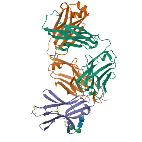
- Additional UniProt keywords include 3D-structure, Adaptive immunity, Glycoprotein, Immunoglobulin domain, Phosphoprotein, and Signal, reflecting its structural and immunological roles.
Experimental Guidance and Technical Tips
- The immunogen corresponds to the extracellular region (amino acids 31–241) of human BTLA, recognized in Western blot and ELISA applications.
- For Western blot, consider using reduced whole cell lysates from human, mouse, or rat lymphocyte lines, given the antibody's cross-reactivity.
- The predicted molecular weight is 33 kDa; however, post-translational modifications such as glycosylation may result in a higher observed band.
- Validate the antibody in your specific sample system and application context, as performance may vary.
CamelBio: Your One-Stop Sourcing Bridge
CamelBio provides diagnostic manufacturers, laboratories, and research institutes with one-stop access to IVD raw materials, technical services, and consulting—from early concept through clinical application. This BTLA antibody, targeting an immune checkpoint receptor, is part of a broad portfolio that includes validated antibody pairs, optimized monoclonal and polyclonal antibodies, bulk ancillary reagents, and custom sourcing for rare targets. We help streamline your immunology and oncology diagnostic development with reliable supply and expert support.
REQUEST A QUOTE
Our professional team will reply to you within one business day. Please feel free to contact us!
Related Articles
Why Direct Hapten Coupling Changes the Economics of Small-Molecule Biosensors
Compare direct hapten coupling with protein conjugates to improve ligand density, signal quality, antibody efficiency, and chip reusability.
From Molecular Recognition to Reliable Results: Engineering Biosensors for Rapid Safety Testing
How antibodies and enzymes turn invisible molecular events into reliable biosensor signals, and how material quality determines test performance.
qPCR Primer Design: The Small Decisions That Determine Assay Reliability
A practical guide to qPCR primer design, from amplicon length and 3' stability to validation for efficient, reproducible diagnostics.
ROX in qPCR: The Quiet Reference That Makes Diagnostic Signals Trustworthy
Learn how ROX normalizes qPCR fluorescence, stabilizes Ct calls, and shapes the trade-off between instrument compatibility and multiplexing.
When a Surface Becomes a Sensor: Choosing Between QCM, SAW, and Six Classes of Quality Detection
Explore six sensor families, how QCM and SAW convert molecular binding into frequency shifts, and why surface chemistry determines diagnostic reliability.
When the Surface Becomes the Assay: Direct Hapten Coating for More Efficient IVD Biosensors
Compare direct hapten derivative coating with protein conjugates to improve SPR assay sensitivity, reduce antibody use, and extend chip life.
The Silent Well: How No Template Controls Protect Molecular Diagnostic Integrity
A practical guide to No Template Controls: how unexpected qPCR signals reveal contamination, reagent impurities, artifacts, and broken workflow controls.
ROX in Real-Time PCR: The Optical Trade-Off Behind Precision and Multiplexing
Understand how ROX normalization corrects qPCR signal variation, how optics consume channels, and how to choose the right master mix.
From Fluorescence to Diagnostic Truth: Choosing Passive Reference Dyes for qPCR Master Mixes
Learn how passive reference dyes stabilize qPCR signals, improve diagnostic reproducibility, and guide ROX formulation across instrument platforms.
Why Minor Groove Binders Turn Short Hydrolysis Probes into Precision Diagnostic Tools
MGB chemistry improves probe stability, SNP discrimination, signal-to-noise ratio, and low-copy detection in demanding molecular assays.
From Molecular Binding to Manufacturing Discipline: How Reliable ELISA Kits Are Engineered
Explore the four operational mechanisms that govern ELISA kit performance, from passive coating and washing to signal generation and scalable IVD manufacturing.
Engineering the Weakest Link: Recombinant Antibodies for Reliable Multi-Analyte Biosensors
Learn how engineered recombinant antibodies improve multi-analyte biosensors through uniform recognition, oriented surfaces, avidity, and wider dynamic range.