The Sample That Looks Like a Number
An ELISA result often arrives as a clean number: 42.6 ng/mL, printed in a report and ready to enter a spreadsheet.
But the sample that produced it may be anything but clean.
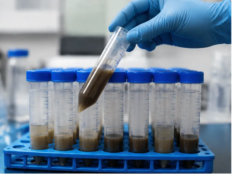
A serum sample contains endogenous proteins, immunoglobulins, salts, and lipids. A plant extract may carry polyphenols and pigments that interact with proteins. Dairy powders introduce fat, proteins, and variable ionic conditions. These components can change how antibodies bind, how enzymes react, and how optical signals develop.
The instrument does not know the difference between a true analyte signal and a matrix-induced signal.
That is why matrix interference should be treated as a design parameter rather than a late-stage defect. By the time a failed recovery study appears during validation, the problem may already be embedded in the antibody pair, buffer system, sample protocol, and calibration model.
Reliable ELISA development begins earlier: by assuming that the matrix will affect the assay and building several defenses against it.
Why Complex Matrices Distort ELISA Performance
An ELISA depends on controlled molecular interactions. The target must remain available. The antibody must recognize it. Non-specific proteins must stay away from the binding surface. The enzyme-substrate reaction must reflect the amount of bound analyte.
A complex matrix can disturb each step.
The result usually appears in one of two forms:
- Signal suppression: the measured concentration is lower than the true concentration.
- Elevated background: non-specific interactions produce a signal that resembles a positive result.
Neither problem is merely technical noise. Both can change clinical interpretation, screening decisions, or research conclusions.
The Psychology of a Plausible Result
The most dangerous assay error is not an obviously failed plate. It is a result that looks reasonable.
A standard curve can appear smooth while the sample matrix changes its slope. A blank can remain low while recovery quietly falls to 60%. An assay may perform well in buffer and fail only when it meets real specimens.
This creates a psychological trap: developers tend to trust visual order. A neat curve feels like evidence of control.
Matrix validation exists to challenge that feeling with controlled experiments.
Diagnose the Matrix Before Trying to Fix It
Interference cannot be managed reliably until it is measured. Two studies provide the first practical map of the problem.
Spike-and-Recovery Testing
Add a known quantity of purified analyte to an unprocessed sample matrix. Then compare the measured increase with the expected increase.
A commonly acceptable recovery range is:
| Result | Likely interpretation |
|---|---|
| 80-120% recovery | Generally acceptable matrix impact |
| Below 80% | Signal suppression, analyte loss, or incomplete extraction |
| Above 120% | Matrix-enhanced binding or elevated non-specific signal |
Recovery should be evaluated across multiple matrix lots, not just one convenient specimen. A single clean sample can conceal the variability that later appears in production or routine testing.
For regulatory-oriented development, a narrower internal target such as 95-107% recovery, with RSD below 2%, provides a stronger basis for confidence when the intended use permits it.
Dilution Linearity
Prepare serial dilutions of the sample in assay buffer. Correct each result for the dilution factor and compare the calculated concentrations.
A regression slope between 0.85 and 1.15 generally indicates acceptable dilutional behavior. Significant deviation suggests that the matrix is changing the binding dynamics as its concentration changes.
Dilution linearity answers a question that spike recovery alone cannot:
Does the assay behave consistently when the matrix is gradually removed?
Together, the two studies help distinguish between analyte loss, signal suppression, non-specific enhancement, and concentration-dependent effects.
A Three-Layer Defense System
Matrix control works best as a layered system.
No single centrifugation step can compensate for a poorly chosen antibody. No blocking reagent can fully rescue an analyte that was lost during aggressive precipitation. No calibration curve can make an unstable sample preparation protocol reproducible.
The most reliable workflow combines:
- Physical and chemical sample cleanup
- Buffer and assay-condition optimization
- Matrix-aware calibration and reagent selection
Each layer addresses a different part of the problem.
Layer One: Remove the Bulk Interferents
The first layer acts before the immunochemical reaction begins. Its purpose is to remove particles, lipids, proteins, pigments, or other components that would otherwise overwhelm the assay.
Centrifugation
Centrifugation is often the least disruptive first step. It removes insoluble particles and can separate some lipid-rich components without exposing the analyte to harsh chemistry.
Its limitation is equally important: centrifugation does not remove every dissolved interferent. A clear supernatant may still contain proteins, salts, polyphenols, or endogenous antibodies that affect the assay.
Filtration and Deproteinization
Membrane filtration can reduce particulates and some high-molecular-weight contaminants. Deproteinization may be useful when endogenous serum proteins create high background, especially in direct ELISA formats.
However, aggressive chemical precipitation can create a second problem. Trichloroacetic acid and similar reagents may co-precipitate lipophilic or low-abundance analytes. The sample may look cleaner while the target has disappeared with the waste fraction.
When analyte recovery is fragile, membrane filtration or affinity-based pre-separation may preserve performance better than harsh precipitation.
Solid-Phase Extraction
Solid-phase extraction, or SPE, can both purify and pre-concentrate trace analytes. This makes it useful for complex plant extracts and other samples in which the target is present at very low levels.
SPE must be developed around the analyte, not selected as a generic cleanup ritual. Important variables include:
- Sorbent chemistry
- Sample loading solvent
- Wash composition
- Elution strength
- Analyte recovery
- Compatibility of the final eluate with the ELISA
Steam distillation may also be appropriate for selected volatile or semi-volatile targets. It should be treated as a target-specific separation method rather than a universal solution for complex matrices.
The Central Trade-Off
Every cleanup step removes something. The question is whether it removes more interference than analyte.
| Cleanup approach | Main benefit | Main risk |
|---|---|---|
| Centrifugation | Low disruption; removes particulates | Limited removal of dissolved interferents |
| Filtration | Simple reduction of solids and aggregates | Adsorption or membrane-related analyte loss |
| Deproteinization | Reduces protein-driven background | Co-precipitation and altered analyte recovery |
| SPE | Purification and pre-concentration | More development variables and operator dependence |
| Steam distillation | Useful for selected volatile targets | Unsuitable for non-volatile or unstable analytes |
The best protocol is often the mildest one that produces acceptable recovery and dilutional behavior.
Layer Two: Use Buffer Chemistry as a Shield
After sample cleanup, residual matrix components remain. Buffer chemistry controls how strongly those components influence the assay.
A few percentage points of surfactant or a small shift in pH can change background, recovery, and apparent affinity. These adjustments should be screened systematically rather than added by intuition alone.
Surfactants and Polymers
Surfactants in the approximate range of 0.1-1.0% may reduce non-specific adsorption and improve wetting. Polymers in the approximate range of 0.1-5% can help block unwanted interactions or stabilize assay components.
The usable concentration depends on the antibody, enzyme label, plate surface, and sample type. Excess surfactant can damage useful interactions or alter the activity of labeled reagents.
Carrier Proteins
Carrier proteins such as BSA can occupy non-specific binding sites and stabilize reagents. They may also introduce new risks if the sample contains anti-animal antibodies or if the carrier interacts with the target.
A carrier protein is therefore part of the assay chemistry, not an invisible background ingredient. Its impact should be evaluated through blank signal, recovery, precision, and cross-reactivity studies.
Neutralizing Plant-Derived Polyphenols
Polyphenols in plant extracts can bind proteins and interfere with enzyme-based detection. Polyvinylpyrrolidone, or PVP, is commonly evaluated as a neutralizing component because it can bind certain polyphenolic compounds.
PVP concentration and extraction conditions must be optimized together. A formulation that suppresses plant-derived interference may also affect analyte extraction or antibody binding if used without control experiments.
pH, Ionic Strength, and Wash Conditions
The assay buffer should provide a stable environment for antibody-analyte binding while limiting non-specific interactions.
Key variables include:
- Sample and assay pH
- Ionic strength
- Blocking reagent composition
- Wash volume and number of cycles
- Incubation time and temperature
- Compatibility between extraction and detection buffers
The goal is not to make the buffer chemically complex. It is to make its behavior predictable across the intended sample range.
Layer Three: Calibrate the Assay for Reality
Even after cleanup and buffer optimization, some matrix effect may remain. Calibration is where the assay acknowledges that reality.
Why Generic Buffer Standards Can Mislead
A standard curve prepared in a clean buffer describes how the assay behaves in that buffer. It does not necessarily describe how the assay behaves in serum, dairy powder, or plant extract.
When standards and samples occupy different chemical environments, the same analyte concentration can produce different signals. The curve may retain an attractive R² while the calculated sample concentration is biased.
Matrix-Matched Calibration
Matrix-matched standards are prepared in a target-free version of the intended sample matrix, or in a validated surrogate that reproduces its relevant behavior.
This approach helps compensate for matrix-induced changes in:
- Antibody binding
- Analyte recovery
- Enzyme activity
- Optical background
- Signal slope
- Detection limits
A well-designed calibration model should support analytical linearity, commonly targeting R² greater than 0.99, while maintaining defensible recovery and precision across the working range.
The difficult part is sourcing a genuinely target-free matrix. For unique patient samples or rare biological materials, this may be expensive or impossible. The surrogate must then be characterized rather than assumed to be equivalent.
Reagent Affinity and Specificity
High-affinity antibodies provide more binding power when matrix components compete with the target. But affinity alone is not enough.
The reagents also need:
- Strong specificity
- Low cross-reactivity
- Stable performance across pH and ionic conditions
- Adequate lot-to-lot consistency
- Compatibility with the chosen assay format
This is where raw-material strategy becomes inseparable from assay design. The antibody, enzyme conjugate, blocking system, and calibrator must be evaluated as a working system.
When the Assay Format Must Change
Sometimes the correct response to matrix interference is not another buffer experiment. It is a different assay architecture.
Direct ELISA
Direct formats can be efficient, but they may be vulnerable to non-specific binding when sample proteins or pigments interact with the detection surface.
Sandwich ELISA
A sandwich format can improve specificity when the analyte is large enough and presents multiple accessible epitopes. Capture and detection antibodies create an additional level of molecular discrimination.
Competitive ELISA
Competitive formats are often better suited to small molecules with limited epitope availability. They can also support analytes for which a conventional sandwich design is structurally impractical.
The format should follow the analyte's molecular size, epitope availability, abundance, and matrix behavior. A familiar format is not necessarily the most robust format.
Manage Wide Concentration Ranges With Bracketed Dilution
Complex samples often contain analyte concentrations that span several orders of magnitude. One dilution rarely provides both sensitivity and reliable quantification.
A bracketed scheme can include dilutions such as:
- 1:10
- 1:70
- 1:610
- 1:4270
The exact series should be adapted to the sample and assay range. The purpose is to create overlapping opportunities for a sample to fall inside the validated linear range.
This approach adds pipetting and data-handling requirements. Automation, flow-injection analysis, or microfluidic preprocessing can reduce manual burden in high-throughput environments, but these systems increase development time and upfront investment.
For lower-throughput laboratories, a carefully validated manual dilution workflow may remain the more practical option.
Match the Strategy to the Product Goal
There is no single optimal interference strategy. The correct balance depends on what the kit must accomplish.
| Development priority | Recommended emphasis | Main caution |
|---|---|---|
| Maximum sensitivity for trace analytes | High-affinity antibodies, gentle cleanup, matrix-matched calibration | Excessive dilution can erase the signal |
| High-throughput screening | Bracketed dilution, streamlined extraction, automated handling | Automation adds cost and validation burden |
| Regulatory-ready accuracy | Multi-lot recovery, dilution linearity, precision, matrix-matched standards | Evidence must cover real intended-use matrices |
| Complex plant extracts | PVP-containing extraction conditions, SPE, unified buffer development | Polyphenol control must not reduce target recovery |
| Lipophilic analytes | Mild separation and recovery-focused extraction | Chemical precipitation may remove the target |
The development team should decide which performance attribute has priority before optimizing individual parameters. Otherwise, every improvement can create a new conflict: cleaner samples but lower recovery, higher sensitivity but more background, or broader range but weaker precision.
Build a Validation Map, Not a Single Pass-Fail Test
Matrix interference varies across lots, operators, seasons, suppliers, and sample preparation histories. A defensible validation plan should reflect that variability.
At minimum, evaluate:
- Multiple representative matrix lots
- Low, mid, and high analyte concentrations
- Spike-and-recovery performance
- Dilution linearity
- Intra-assay and inter-assay precision
- Blank and background signal
- Cross-reactivity
- Stability through extraction and storage
- Lot-to-lot performance of critical reagents
A useful validation record connects each observed failure to a corrective action. Low recovery may require extraction changes. Elevated background may require blocking or wash optimization. Non-linearity may require dilution, calibrator redesign, or a format change.
This turns troubleshooting from a sequence of guesses into an engineering process.
Matrix Interference Is a Controllable Variable

The matrix is part of the assay environment. It cannot always be removed, and it should not be treated as an afterthought.
A reliable ELISA for complex samples is built through coordinated decisions:
- Remove bulk interferents with the least disruptive preparation method.
- Tune buffer chemistry to control residual non-specific interactions.
- Select antibodies and assay formats that remain effective under real conditions.
- Calibrate in a matrix that represents the intended sample.
- Validate recovery, dilution behavior, precision, and linearity across relevant lots.
CamelBio supports diagnostic manufacturers, laboratories, and research institutes with one-stop access to IVD raw materials, technical services, and consulting across the path from concept to clinic. For assay teams working through antibody selection, blocking reagents, buffer formulation, calibrator design, or broader development decisions, Contact Our Experts to build matrix control into the assay from the beginning.
Related Products
- Anti-Emerin/EMD Polyclonal Antibody for WB, IHC-P, IF/ICC, ELISA - P50402
- Anti-Olig2 Rabbit Monoclonal Antibody for WB, ELISA - Q13516
- Anti-Alpha-Fetoprotein (AFP) Monoclonal Antibody for WB, IF/ICC, ELISA - P02771
- Anti-STX1A Polyclonal Antibody for WB, IF/ICC, ELISA - Q16623
- Anti-Cyclin E2 Monoclonal Antibody for WB, ELISA - O96020

![Anti-eIF4E Rabbit Monoclonal Antibody [KD Validated] for WB, IF/ICC, IP, ELISA - P06730](https://image.camelbiological.com/images/products/4940/main_image_6a636cf2795024.14314593-small.webp)