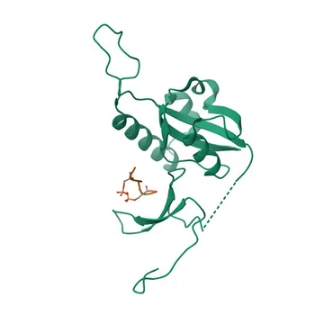
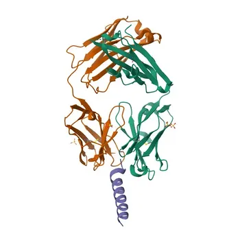
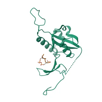
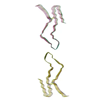
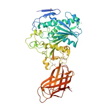
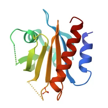
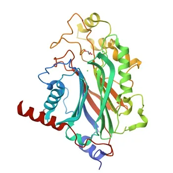
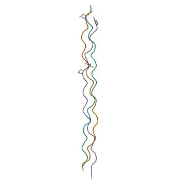
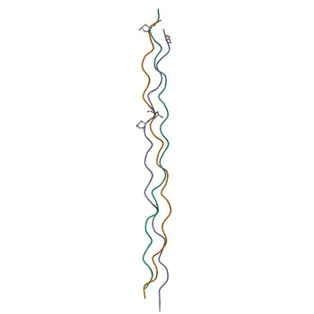
While the core amino acid sequence of Procollagen Type I N-Terminal Propeptide (PINP) is identical in all its forms, the critical structural difference engineers must confront is quaternary organization. In healthy circulation, PINP is a stable homotrimeric molecule held together by non-covalent interactions between three identical chains. However, in specific disease states like end-stage renal disease (ESRD) or prolonged immobilization, degraded monomeric fragments of PINP accumulate in plasma. This single structural dichotomy—intact trimer versus monomeric fragment—directly dictates the entire design philosophy behind an “intact” versus a “total” PINP immunoassay.
The decision to measure only the trimeric PINP or both trimeric and monomeric forms is not a nuance; it defines antibody specificity, calibration material composition, and ultimately the assay’s clinical sensitivity across patient populations. An intact assay must recognize a quaternary epitope unique to the trimer, while a total assay must capture a linear epitope that survives fragmentation.
The Structural Landscape of Circulating PINP
The Native State: A Stabilized Homotrimer
The propeptide released during type I collagen synthesis is an exceptionally stable, non-covalently associated trimer. This quaternary structure is the predominant form in healthy individuals and is directly proportional to the amount of newly synthesized collagen fibrils.
The Degradative Shift: Monomeric Fragments in Disease
In populations with impaired renal clearance or prolonged recumbency, the trimeric PINP is partially degraded into smaller monomeric chains. These monomers accumulate because they are not filtered and cleared as efficiently as the intact trimer.
This structural shift means that a blood sample from an ESRD patient contains a heterogeneous mix of PINP species—the intact trimer alongside its monomeric breakdown products. An immunoassay’s ability to differentially detect these forms determines its diagnostic accuracy.
Engineering the Intact PINP Immunoassay
Antibody Target: A Quaternary Conformational Epitope
To measure only the intact trimer, the capture or detection antibody must recognize an epitope that exists only when the three chains are associated. This epitope is typically a structural motif created by the apposition of two or more monomeric subunits at the trimer interface.
A purely linear peptide immunogen will fail to generate these antibodies. Successful development requires immunizing with a purified, stabilized trimeric PINP to elicit a response against the native quaternary folds.
Risk of Cross-Reactivity and the Calibration Standard
The calibrator must be a pure, properly folded intact PINP trimer. If the calibration material contains even trace amounts of monomer, the assay’s sensitivity for the trimer will be overestimated, leading to inaccurate low-end quantitation.
This format achieves maximal specificity for new collagen synthesis, as the trimer is the immediate product of procollagen cleavage. It explicitly ignores the monomeric pool that does not directly reflect instantaneous bone formation.
Engineering the Total PINP Immunoassay
Antibody Target: A Linear, Cryptic-Resistant Epitope
A total assay requires a pair of monoclonal antibodies that bind a linear peptide sequence present on both the intact trimer and its free monomeric fragments. The epitope must be fully accessible in the folded trimer and not sterically hidden at the subunit interface.
Immunization can utilize a synthetic peptide of the PINP sequence. However, rigorous screening is mandatory to ensure the chosen antibody pair demonstrates equimolar reactivity—identical binding affinity whether the analyte is in trimeric or monomeric form—to avoid systematic bias.
The Calibrator Challenge: Mimicking the Disease-State Matrix
Calibrating a total assay is uniquely demanding. The ideal standard is not pure trimer alone, but a commutable material containing both trimeric and monomeric PINP in a defined ratio that mirrors the intended clinical population.
Using a trimer-only standard will incorrectly spike the dose-response curve and cause under-recovery of samples rich in monomers. A total PINP assay is inherently a summation measurement and must be validated with samples known to contain high fragment loads.
Understanding the Trade-offs
Clinical Specificity Versus Population Inclusivity
An intact PINP assay provides a direct, unconfounded readout of bone formation activity in patients with normal glomerular filtration rates. However, in renal failure, where the monomeric pool expands dramatically, the intact assay may underestimate the total PINP-related signal, potentially missing valuable prognostic information about the overall collagen turnover burden.
A total PINP assay captures the entire molecular landscape, which is advantageous in monitoring patients on hemodialysis or bedridden elderly individuals where fragment accumulation is the dominant variable. Yet this inclusivity comes at a cost: the detected signal now conflates freshly produced trimer with renally retained degradation products that may not dynamically reflect instantaneous changes in bone formation.
Pre-Analytical Stability: A Shared Advantage
Importantly, the structural robustness noted in the supplementary references applies to both forms. PINP—whether trimeric or monomeric—exhibits exceptional stability in serum and plasma at room temperature for up to one week. This means that once a sample is drawn, the ratio of trimer to monomer does not artifactually shift due to ex vivo degradation, making both assay formats analytically robust in logistics.
Common Pitfalls to Avoid
- Antibody blindspot in intact assays: Using an antibody that binds a linear epitope easily accessible in the monomer but partially occluded in the trimer. This creates a “total-like” assay that fails its intended specific measurement.
- Improper calibrator for total assays: Using a recombinant trimer standard and assuming it will calibrate monomeric species identically, leading to gross inaccuracy when fragments dominate the sample.
- Ignoring the patient population mix: Developing an intact assay for an osteoporosis clinic that unknowingly includes a significant subset of patients with chronic kidney disease stage 4, resulting in misleadingly low biomarker values.
Making the Right Engineering Choice for Your Clinical Goal
The decision hinges on the specific patient cohort and the clinical question the assay is meant to answer.
- If your primary focus is monitoring anabolic or antiresorptive therapy in generally healthy postmenopausal osteoporosis patients: Choose the intact PINP format. It provides the purest, most responsive biomarker of new collagen synthesis, unaffected by renal clearance artifacts.
- If your primary focus is managing bone disease in end-stage renal disease or assessing total skeletal turnover in bedridden, multi-morbid elderly patients: A total PINP assay is necessary to capture the clinically relevant, fragment-heavy analyte pool. Accept that the measurement represents a composite of production and retention, requiring population-specific reference intervals.
- If your goal is a universal, broad-use kit intended for mixed hospital and specialty labs: Total PINP offers practical inclusivity, but you must heavily invest in demonstrating equimolar mono/tri reactivity and provide clear instructions for use, explicitly warning that the signal in renally impaired patients partly reflects impaired clearance, not solely formation.
Your engineering choice transforms a simple molecular difference into a tool either exquisitely precise or broadly vigilant—always align the assay’s structural specificity with the biological reality of the patient you aim to serve.
Summary Table:
| Parameter | Intact PINP Immunoassay | Total PINP Immunoassay |
|---|---|---|
| Target Analyte | Native intact homotrimer | Homotrimer + monomeric fragments |
| Epitope Type | Quaternary (conformational interface) | Linear (cryptic-resistant sequence) |
| Calibrator Requirement | Pure, properly folded intact PINP trimer | Commutable standard with equimolar reactivity |
| Ideal Patient Population | Osteoporosis / normal renal clearance | ESRD, hemodialysis, elderly/bedridden cohorts |
| Renal Function Sensitivity | Unconfounded by renally retained monomers | Reflects combined synthesis and clearance accumulation |
Accelerate Your PINP Immunoassay Development with CamelBio
Engineering precise intact or total PINP assays requires high-affinity antibodies and rigorously characterized calibrators to guarantee clinical accuracy across target patient cohorts.
CamelBio provides diagnostic manufacturers, labs, and research institutes with one-stop access to high-quality IVD raw materials, technical services, and expert consulting—covering every stage from concept to clinic.
- Specific Antibody Pairs: Access antibodies tailored for quaternary trimer epitopes or linear, equimolar monomer/trimer recognition.
- Standardization Support: Source reliable, well-characterized antigens and calibrator matrices to eliminate matrix bias.
- Turnkey Development Consulting: Leverage our expert assay development guidance to fast-track your kit to market.
Ready to elevate your bone biomarker portfolio? Contact CamelBio today to connect with our technical team and request product samples!
Related Products
- Anti-Human Pro-Collagen I alpha (COL1A1) Rabbit Polyclonal Antibody for WB, IF-P, IHC-P - P02452
- Anti-Collagen I/COL1A1 Rabbit Monoclonal Antibody for WB, IHC-P, IF-P, ELISA - P02452
- Anti-Collagen I/COL1A1 Polyclonal Antibody for WB, IHC-P, IF/ICC, ELISA - P02452
- Anti-Collagen I/COL1A2 Monoclonal Antibody for WB, ELISA - P08123
- Anti-Collagen I/COL1A1 Rabbit Polyclonal Antibody for WB, IF/ICC, IHC-P, ELISA - P02452
![[KD Validated] Pin1 Rabbit mAb for WB and ELISA - Q13526](https://image.camelbiological.com/images/products/3649/main_image_6a63526aefb330.70106281-small.webp)