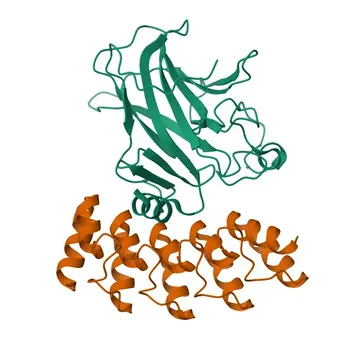
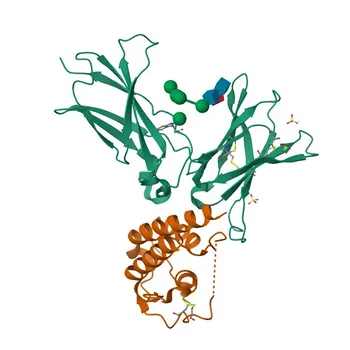
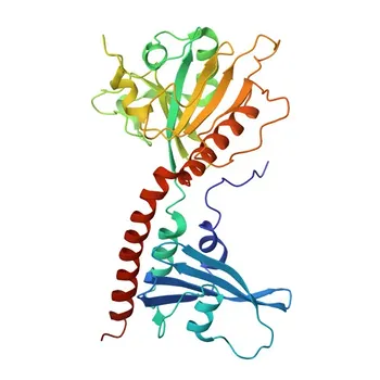
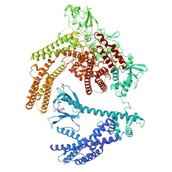
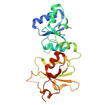
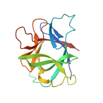
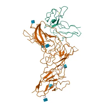
The answer lies in a fundamental dichotomy of protein architecture. Linear epitopes are continuous sequences of 6–15 amino acids (or 2–7 sugar residues) recognized in their primary structure. Conformational epitopes are formed by discontinuous residues brought into spatial proximity by the protein’s folded 3D shape. This structural distinction is the decisive factor for antibody screening in diagnostic assays: you must select antibodies using a target that presents the same epitope state—native or denatured—that will exist in your final test system. Failure to match them leads to poor binding, false negatives, and lost assay sensitivity.
Epitope type dictates not just what an antibody sees, but how robustly and under what conditions it sees it. For reliable assay performance, antibody screening must faithfully replicate the structural state of the target protein expected in your diagnostic workflow—native conformational integrity or denatured linear exposure.
The Fundamental Architecture of Epitopes
Every antibody-binding site on an antigen falls into one of two structural categories. Understanding their molecular basis is the first step to designing a rational screening strategy.
Linear Epitopes: Continuous Recognition Sites
A linear epitope is a continuous stretch of monomeric units—typically 6 to 15 amino acids or 2 to 7 sugar residues—along the polypeptide chain.
Because recognition relies solely on the primary sequence, these epitopes remain accessible even if the protein unfolds or denatures.
They are often internal segments that become exposed only when the native structure is disrupted.
Conformational Epitopes: The Role of 3D Folding
A conformational epitope consists of non-contiguous amino acids that are widely separated in the sequence but brought together by secondary and tertiary folding.
Their structure depends on the delicate spatial arrangement of the protein’s native state.
Any alteration that disrupts this folding—heat, detergents, reduction, or pH extremes—can permanently destroy the epitope and abolish antibody binding.
Why Epitope Structure Dictates Antibody Screening Strategies
Screening for diagnostic antibodies is not just about finding a binder; it is about finding one that works in the exact environmental and processing conditions of your final assay. The epitope type shapes every decision.
Assay Formats and Target State: Matching the Right Antibody
In denaturing assays (e.g., Western blotting, certain sample-preparation steps), proteins are reduced and unfolded.
Here you must screen with antibodies that recognize linear epitopes, because native conformational targets no longer exist.
Conversely, native-state liquid-phase platforms—such as automated chemiluminescent immunoassays (CLIA), sandwich ELISAs, or immunoturbidimetric tests—require antibodies optimized for intact conformational epitopes on the folded protein.
The Impact of Denaturation and Sample Processing
Chemical fixation, reduction, aggregation, matrix masking, and shifts in pH or solvent can dramatically alter epitope availability.
Conformational epitopes are destroyed if sample preparation denatures the protein; screening antibodies against native antigen will yield binders that fail in the processed sample.
Linear epitopes, on the other hand, might be newly exposed upon unfolding, making them the correct target for assays where denaturation is unavoidable.
Practical Considerations for Antibody Selection and Reagent Optimization
Beyond the assay format, epitope characteristics influence buffer formulation, antibody pairing, and raw material stability.
Buffer Tolerance and Epitope Stability
Antigens presenting linear epitopes are robust and can tolerate vigorous detergents and a wide pH range without losing reactivity.
Conformational epitopes are fragile: high salt, aggressive detergents, or incorrect pH can disrupt the tertiary structure, restricting your choice of buffer components.
During antibody screening, you must replicate the final sample diluent and reaction buffers to ensure the antibody’s binding profile remains consistent.
Avoiding Steric Hindrance in Sandwich Assays
When selecting antibody pairs for sandwich-format assays, the spatial orientation of epitopes matters.
If both capture and detection antibodies target overlapping or closely spaced conformational epitopes, steric hindrance can prevent sandwich complex formation.
Screening panels that include antibodies to distinct, non-overlapping epitopes—whether linear or conformational—preserves assay signal and dynamic range.
Understanding the Trade-offs
No single epitope type is universally superior. The choice involves clear compromises that must be weighed against your diagnostic requirements.
Linear Epitopes: Robust but Potentially Less Specific
Linear epitopes survive harsh processing, making them ideal for robust, reproducible assays.
However, their simplicity can lead to higher cross-reactivity with homologous sequences in other proteins, potentially reducing specificity.
They may also be buried in the native protein, making them invisible in native-state assays unless purposely exposed.
Conformational Epitopes: High Specificity, High Fragility
Conformational epitopes often confer exquisite specificity because they rely on the unique folding of a single protein.
Yet this very specificity makes them exquisitely sensitive to minor changes in buffer composition, temperature, or sample handling.
Screening with native protein in a physiological buffer is mandatory; any deviation can select antibodies that later fail in production.
The Screening Dilemma: Native vs. Denatured Antigen Selection
If your final assay uses a denatured target, you must screen with a denatured or peptide antigen—antibodies raised against a native protein will likely miss the linear epitopes that dominate the processed sample.
Conversely, screening with only peptide or denatured protein will yield antibodies that cannot bind native targets in a liquid-phase sandwich assay.
The most common failure in antibody sourcing is mismatch between the screening antigen’s conformation and the diagnostic target state.
Making the Right Choice for Your Diagnostic Assay
Align your antibody screening approach with the specific demands of your assay platform and the target’s expected structural state.
- If your primary focus is a denaturing or sample-pretreatment assay: Screen candidate antibodies against linear epitopes—use peptide libraries or denatured protein to ensure reactivity survives the processing steps.
- If your primary focus is a native-state liquid-phase platform: Screen with intact, native protein in a buffer that mimics the final sample diluent; prioritize antibodies that bind conformational epitopes and validate their stability under realistic handling conditions.
- If your primary focus is a sandwich assay using raw materials from multiple vendors: Map epitope specificity early to avoid steric clashes—select capture and detection antibodies that recognize topographically distinct epitopes, whether linear or conformational.
A truly optimized diagnostic assay emerges when you treat epitope structure not as an academic nuance, but as the blueprint for antibody selection.
Summary Table:
| Feature | Linear Epitopes | Conformational Epitopes |
|---|---|---|
| Structural Basis | Continuous amino acid sequence (6–15 AAs) | Discontinuous residues folded into 3D proximity |
| Target Protein State | Unfolded / Denatured | Intact / Native 3D Fold |
| Environmental Robustness | High; resists heat, detergents, and pH extremes | Low; fragile to denaturation, heat, and pH shifts |
| Performance Profile | Robust exposure; higher risk of cross-reactivity | Exquisite specificity; risk of loss-of-binding if denatured |
| Recommended Assay Formats | Western blot, denaturing pretreatment assays | CLIA, Sandwich ELISA, Immunoturbidimetric assays |
Developing a new diagnostic assay and need the right antibody pairs for your platform? Whether your workflow relies on denatured targets or native 3D conformational states, CamelBio provides diagnostic manufacturers, labs, and research institutes with one-stop access to premium IVD raw materials, technical services, and expert consulting—covering every stage from concept to clinic.
Avoid costly screening mismatches and ensure peak assay sensitivity. Contact CamelBio today to optimize your antibody selection and streamline your diagnostic development!
Related Products
- Anti-p63 Rabbit Monoclonal Antibody for WB, ELISA, ChIP - Q9H3D4
- Anti-DET1 Polyclonal Antibody for WB and ELISA - P48732
- Anti-STX1A Polyclonal Antibody for WB, IF/ICC, ELISA - Q16623
- Anti-Human/Monkey IgD Monoclonal Antibody for Flow Cytometry - P01880
- Anti-IL21 Rabbit Monoclonal Antibody for WB and ELISA - Q9HBE4