Your antigen selection strategy for T. b. gambiense immunoassays must start with the immunodominant VSG LiTat 1.3, but its diagnostic blind spots—false negatives from gene-negative strains and false positives in malaria-endemic populations—demand a more robust, multi-target approach. A raw material design that combines multi-epitope recombinant antigens with optimized diluents can overcome these limitations, delivering assays that capture broader strain variability without sacrificing specificity.
The central challenge is balancing high sensitivity with reliable specificity. LiTat 1.3 provides a strong foundation, yet developers must address its strain-dependent gaps and serological cross-talk to create a rapid test that works across diverse geographical and genetic backgrounds.
The Central Role of VSG LiTat 1.3 and Its Limits
The variant surface glycoprotein LiTat 1.3 is the most widely validated serological target, but relying on it alone introduces actionable risks.
Why LiTat 1.3 Is the Gold Standard
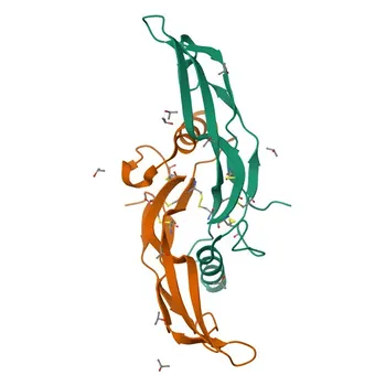
Serological screening for Trypanosoma brucei gambiense consistently leverages the VSG LiTat 1.3 due to strong antibody responses in infected individuals. In well-characterized assays, it can achieve sensitivities between 87% and 98%, making it a cornerstone antigen for card agglutination and immunoassay kits. Its immunodominance simplifies raw material selection and historical validation data are abundant.
The Risk of False Negatives from Strain Variability
The major pitfall is a false-negative result in patients infected with parasite strains that lack the LiTat 1.3 gene. These gene-negative variants circulate in certain endemic foci, and a single-antigen test will miss them entirely. Diagnostic developers must therefore account for this genetic heterogeneity at the raw material stage, as it directly impacts clinical sensitivity in the field.
Navigating Cross-Reactivity in Endemic Settings
Population-specific immunological noise can erode specificity. The primary concern is serological cross-reactivity with malaria, a co-endemic infection that generates polyclonal antibodies capable of binding LiTat 1.3 non-specifically.
Malaria-Induced False Positives
In malaria-endemic populations, even a well-designed LiTat 1.3-based assay can yield false-positive results. This happens because chronic malaria exposure drives production of cross-reactive antibodies that recognize parasitic epitopes shared with Plasmodium species. For a screening tool meant for rural clinics, such false positives not only misdiagnose individuals but also undermine trust in the test.
Strategies to Reduce Non-Specific Binding
Optimizing immunoassay diluents is a direct countermeasure. By incorporating blocking agents, chaotropic salts, or other proprietary additives, developers can disrupt low-affinity, non-specific interactions with cross-reactive antibodies. This preserves the signal from high-affinity anti-VSG antibodies, effectively raising the signal-to-noise ratio without changing the antigen itself.
Optimizing Raw Materials for Broader Coverage
Addressing the dual challenge of strain escape and cross-reactivity requires moving beyond a single native antigen.
Multi-Epitope Recombinant Antigens
Evaluating multi-epitope recombinant antigens lets you combine immunodominant regions from LiTat 1.3 with conserved epitopes from other VSG variants or invariant antigens. This broadens the capture net, reducing the chance that a parasite lacking LiTat 1.3 will go undetected. Recombinant production also allows precise control over antigen purity and lot-to-lot consistency, critical for IVD manufacturing.
Advanced Immunoassay Diluents
Optimized diluents do more than merely reduce non-specific binding. They can shape the immunological environment to favor detection of high-avidity antibodies, which are more likely to be specific. By fine-tuning the buffer system, protein stabilizers, and surfactants, you shift the assay’s equilibrium toward true positives while suppressing the noise from co-endemic infections like malaria.
Understanding the Trade-offs in Antigen Design
No single formulation will perfectly satisfy all performance metrics. Developers must make informed trade-offs based on the intended use setting.
- Sensitivity vs. Specificity: Adding more epitopes can increase sensitivity by catching diverse strains, but each extra epitope introduces a risk of new cross-reactive targets. You may need to sacrifice a few points of specificity to gain crucial strain coverage.
- Cost and Complexity: Multi-epitope recombinant proteins are more expensive to produce and purify than native LiTat 1.3. For a rapid diagnostic test intended for low-resource settings, this cost must be weighed against the public health cost of missed infections.
- Validation Burden: Each new antigen or diluent component demands rigorous testing across panels from multiple geographic regions, including well-characterized malaria-positive and LiTat 1.3-negative trypanosomiasis cases. The development timeline can stretch as a result.
Making the Right Choice for Your Development Goal
Your raw material selection should be driven by the epidemiological context and the public health priority of your screening program.
- If your primary focus is maximum sensitivity for outbreak detection: Prioritize a multi-epitope recombinant that covers the most common LiTat 1.3-negative variants, even if it means accepting a slight rise in false-positive rates.
- If your primary focus is eliminating false positives in malaria-endemic cohorts: Invest heavily in optimized diluent chemistry to suppress cross-reactive antibodies, and consider a more conservative antigen design centered on LiTat 1.3 with rigorous malaria-panel validation.
- If your primary focus is developing a reference-standard assay for clinical labs: Combine both strategies—a well-characterized multi-epitope recombinant plus a thoroughly formulated diluent—and perform extensive analytical specificity testing to lock in reproducibility across diverse sample matrices.
- If your primary focus is cost-sensitive point-of-care deployment: Start with a high-quality native or recombinant LiTat 1.3, but mandate in-field surveillance to quickly flag regions where gene-negative strains might cause diagnostic failure.
The most successful immunoassay raw materials emerge from this deliberate balance: they do not merely replicate the historical gold standard but proactively engineer around its known vulnerabilities.
Summary Table:
| Development Factor / Challenge | Diagnostic Impact | Recommended Raw Material Strategy |
|---|---|---|
| VSG LiTat 1.3 Gene-Negative Strains | Risk of false negatives in endemic regions lacking the LiTat 1.3 gene | Incorporate multi-epitope recombinant antigens with conserved epitopes |
| Malaria Cross-Reactivity | False positives caused by polyclonal cross-reactive antibodies | Optimize diluents with blocking agents, chaotropic salts, and stabilizers |
| Sensitivity vs. Specificity Trade-off | Broader strain coverage can increase background noise | Fine-tune buffer systems and balance antigen combinations for the target setting |
| Production & Validation Burden | Inconsistency or complex multi-antigen purification | Shift to recombinant expression for lot-to-lot consistency and scalability |
Accelerate your diagnostic development with robust, high-performance raw materials. CamelBio provides diagnostic manufacturers, labs, and research institutes with one-stop access to IVD raw materials, technical services, and consulting—covering every stage from concept to clinic. Whether you are developing rapid screening tests or overcoming cross-reactivity challenges in complex sample matrices, our team is ready to assist. Contact us today to partner on your next immunoassay project!
Related Products
- Anti-TBR1 Monoclonal Antibody for WB, IF-P, ELISA - Q16650
- Anti-p63 Rabbit Monoclonal Antibody for WB, ELISA, ChIP - Q9H3D4
- Anti-PBR/TSPO Monoclonal Antibody for WB - P30536
- Anti-Tau Polyclonal Antibody for WB, IHC-P, IF/ICC, ELISA - P10636
- Anti-tPA/PLAT Rabbit Monoclonal Antibody for WB, IF/ICC, IF-P, ELISA - P00750