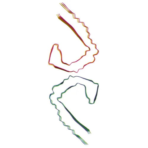
Polyclonal Antibodies
Anti-Tau Polyclonal Antibody for WB, IHC-P, IF/ICC, ELISA - P10636
Item Number : CM0016298
Price varies based on specs and customizations
- Application
- WB, IHC-P, IF/ICC, ELISA
- Cross Reactivity
- Human, Mouse, Rat
- Protein Weight
- 79kDa

Shipping:
Contact us to get shipping details Enjoy On-time Dispatch Guarantee.
Why Choose Us
Easy ordering process, quality products, and dedicated support for your business success.
Core Product Specifications and Parameters
| Parameter | Value |
|---|---|
| Product Name | Tau Rabbit pAb |
| Remarks/Alias | TAU; MSTD; PPND; DDPAC; MAPTL; MTBT1; MTBT2; tau-40; FTDP-17; PPP1R103; Tau-PHF6; Tau |
| Species | Human |
| Gene ID (Human) | 4137 |
| Gene ID | 4137 |
| Immunogen | A synthetic peptide corresponding to a sequence within amino acids 658-758 of human Tau (NP_058519.3) |
| Source | Rabbit |
| Category | Polyclonal Antibodies |
| Application | WB, IHC-P, IF/ICC, ELISA |
| Cross Reactivity | Human, Mouse, Rat |
| SWISS | P10636 |
| Protein Weight | 79kDa |
| Shipping | Ice bag |
Biological Background: Tau Function and Localization
- Tau (Microtubule-associated protein tau) promotes microtubule assembly and stability and is involved in the establishment and maintenance of neuronal polarity. Related references: PMID:21985311
- Tau acts as a linker protein, with its C-terminus binding axonal microtubules and its N-terminus binding neural plasma membrane components. Related references: PMID:21985311 PMID:32961270
- Axonal polarity is determined by TAU/MAPT localization near the centrosome; short tau isoforms provide cytoskeletal plasticity, while longer isoforms preferentially stabilize microtubules.
- Expressed primarily in neurons; a peripheral nervous system-specific isoform (PNS-tau) is expressed in the PNS, while other isoforms are central nervous system-specific.
- Subcellular localization includes cytoplasm, cytosol, cell membrane, cytoskeleton, axons, dendrites, and secretion.
- Post-translational modifications include phosphorylation, acetylation, glycation, methylation, O-glycosylation, and ubiquitination; mutations and hyperphosphorylation are linked to Alzheimer disease, Parkinsonism, and other tauopathies.
Experimental Guidance and Technical Tips
- The immunogen spans amino acids 658–758 of human Tau, a C-terminal region; this design may detect multiple isoforms due to alternative splicing.
- In Western blot, expect a primary band near 79 kDa, though post-translational modifications (e.g., phosphorylation) and splice variants could yield additional bands.
- For immunohistochemistry and immunofluorescence, validate in appropriate neuronal tissue or cell models; cross-reactivity with mouse and rat supports use in rodent studies.
- Consider ELISA for quantitative analysis, but note that epitope accessibility may vary with sample preparation.
CamelBio: Your One-Stop Sourcing Bridge
CamelBio provides diagnostic manufacturers, labs, and research institutes with one-stop access to IVD raw materials—from validated antibody pairs and optimized monoclonal/polyclonal antibodies to bulk ancillary reagents. Our portfolio supports neurodegeneration research, including tau biology and Alzheimer’s disease biomarkers. We also assist in sourcing rare raw materials, streamlining your transition from concept to clinic.
REQUEST A QUOTE
Our professional team will reply to you within one business day. Please feel free to contact us!
Related Articles
From Hapten to Reliable Result: Engineering Small-Molecule Immunoassays for Diagnostic Use
Discover how hapten chemistry, antibody selection, competitive formats, and validation determine small-molecule immunoassay performance.

Engineering High-Sensitivity Electrochemical Immunosensors for Small-Molecule Detection
How competitive assay design, high-affinity antibodies, HRP labels, and interface control enable reliable trace small-molecule detection.
Choosing the Right High-Throughput Toxicology Platform: Cell-Based Bioassays Versus ELISA
Compare cell-based bioassays and ELISA for toxicology screening, from biological relevance and throughput to validation and technical support.