The Design Problem Begins with Molecular Geometry
A sensor developer can spend weeks improving electrode conductivity, only to discover that the assay architecture itself has imposed a hard sensitivity limit.
This often happens when a familiar protein-assay strategy is applied to a small molecule.
A sandwich immunoassay works because a protein usually presents several accessible epitopes. One antibody captures the target. A second antibody binds to another region and creates the measurable sandwich.
Small molecules do not offer that geometry.
A small molecule or hapten may be completely enclosed by a single high-affinity antibody. Once that binding event occurs, there is no second surface large or distinct enough for another antibody to recognize. The problem is not a weak electrode or an insufficiently active enzyme. The problem is physical access.
That constraint determines the central design decision:
High-sensitivity electrochemical immunosensors for small molecules must generally use a competitive assay format rather than a sandwich format.
Why Competitive Assays Are Structurally Necessary
In a competitive immunosensor, the sample analyte competes with an immobilized version of the analyte for a limited quantity of labeled antibody.
A typical system contains four functional elements:
- A target molecule in the sample.
- A hapten-protein conjugate immobilized on the electrode.
- An enzyme-labeled monoclonal antibody.
- An electrochemical substrate system that converts enzyme activity into current.
The surface-bound conjugate acts as the fixed competitor. When the sample contains little or no target, more labeled antibody binds to the electrode. The enzyme generates a stronger signal.
When the sample contains a higher concentration of target, the antibody is occupied in solution and is less available to bind to the electrode. The measured signal therefore decreases.
The assay does not follow the intuitive pattern in which more analyte produces more signal.
| Sample target concentration | Labeled antibody bound to electrode | Electrochemical signal |
|---|---|---|
| Low or zero | High | High |
| Moderate | Reduced | Reduced |
| High | Low | Low |
This inverted response is more than a calibration detail. It changes how sensitivity must be engineered.
The Inverted Calibration Curve Changes the Risk Profile
In a conventional direct assay, developers often focus on measuring a signal that rises above the blank.
In a competitive assay, the blank produces the maximum signal. The analytical challenge is detecting a small reduction from that high-signal state.
That means background variation becomes especially damaging. A small amount of nonspecific adsorption, electrode drift, reagent degradation, or matrix interference can resemble the signal change caused by a low concentration of analyte.
The limit of detection is therefore governed by two linked objectives:
- Generate a strong and reproducible maximum signal.
- Reduce every source of variation that could hide a small signal decrease.
This is why signal amplification alone rarely solves the problem. A louder instrument does not help if the background is equally loud.
The Antibody Sets the Biological Performance Ceiling
The most important raw material is the receptor.
For competitive binding, antibody affinity directly influences how effectively the sensor can distinguish very low concentrations of free analyte from the immobilized competitor. The equilibrium dissociation constant, or KD, provides a useful framework for understanding this relationship.
A lower KD generally indicates tighter binding. A high-affinity antibody can maintain a meaningful competitive response at lower analyte concentrations, particularly when the assay is carefully balanced around antibody concentration, competitor density, and incubation time.
But affinity is not the only selection criterion.
A development team should evaluate:
- Association rate.
- Dissociation rate.
- Specificity for the parent compound.
- Cross-reactivity with metabolites and structural analogues.
- Stability under storage and assay conditions.
- Compatibility with labeling chemistry.
- Performance in the intended sample matrix.
The dissociation rate deserves particular attention. Two antibodies can show similar equilibrium affinity while behaving differently during the practical timescale of an assay. An antibody with a slower off-rate may preserve a stronger and more stable immune complex, while an antibody with a faster off-rate can produce greater sensitivity to timing and washing conditions.
The receptor is not simply a binding reagent. It is the kinetic engine of the assay.
Screening Antibodies as an Engineering Program
Antibody screening should begin with the intended measurement problem rather than with a generic ranking of clones.
A clone that performs well in buffer may fail in serum. A clone with excellent parent-compound specificity may bind a clinically irrelevant metabolite. A highly affine antibody may create an assay that is difficult to displace within a practical incubation period.
A more useful screening workflow asks several questions at once:
| Screening question | Why it matters |
|---|---|
| How tightly does the antibody bind the target? | Defines the potential lower detection range |
| How quickly does it associate? | Affects time to equilibrium |
| How slowly does it dissociate? | Influences wash stability and assay reproducibility |
| What analogues does it recognize? | Determines analytical specificity |
| Does labeling alter its binding behavior? | Protects the original receptor performance |
| Does it remain functional in real matrices? | Connects proof of concept to clinical use |
This is where raw-material access and technical interpretation become inseparable. The best clone is not necessarily the one with the most impressive isolated binding number. It is the one that produces the most reliable competitive response in the final assay environment.
Designing the Hapten-Protein Conjugate
The immobilized competitor determines what the antibody sees at the electrode interface.
A common approach links the target hapten to a carrier protein such as bovine serum albumin. The carrier provides a larger structure that can be adsorbed or chemically attached to the solid phase, while the hapten presents the relevant molecular features for antibody recognition.
The conjugate must be designed with control over several variables:
- Hapten orientation.
- Linker length and chemistry.
- Degree of substitution.
- Carrier-protein integrity.
- Surface immobilization behavior.
- Lot-to-lot composition.
The orientation of the hapten can change the apparent epitope presented to the antibody. A linker attached at the wrong position may hide the structural feature that the antibody must recognize. A conjugate with excessive or heterogeneous hapten loading may create a crowded surface with unpredictable binding behavior.
This is why the conjugation ratio should be characterized rather than assumed.
An inconsistent conjugation process changes the effective competitor concentration on the electrode. The result can be a shifted calibration curve, altered dynamic range, and greater variation between production lots.
For a research experiment, this may be an inconvenience. For an IVD product, it becomes a manufacturing risk.
HRP Turns Binding Events into Measurable Electrons
The enzyme label is responsible for converting molecular recognition into an electrical readout.
Horseradish peroxidase is widely used for this purpose because it combines high catalytic activity with established compatibility with electrochemical detection systems. In the presence of hydrogen peroxide and a suitable redox mediator or substrate, HRP can produce a measurable current through repeated catalytic turnover.
The principle is straightforward:
- More HRP-labeled antibody retained on the electrode produces more catalytic activity.
- More catalytic activity produces a stronger current.
- More target in the sample displaces the labeled antibody.
- Displacement reduces the current.
A single binding event can therefore influence many electron-transfer events. This is the source of enzymatic amplification.
However, the label must be optimized as part of the antibody conjugate. Excessive labeling can reduce antibody affinity, increase steric hindrance, or destabilize the conjugate. Insufficient labeling may produce a signal that is too weak for reliable discrimination.
The goal is not maximum enzyme loading in isolation. It is the best balance between:
- Preserved antibody binding.
- High specific activity.
- Storage stability.
- Low nonspecific adsorption.
- Reproducible electrochemical response.
The Electrode Is an Interface, Not Just a Conductor
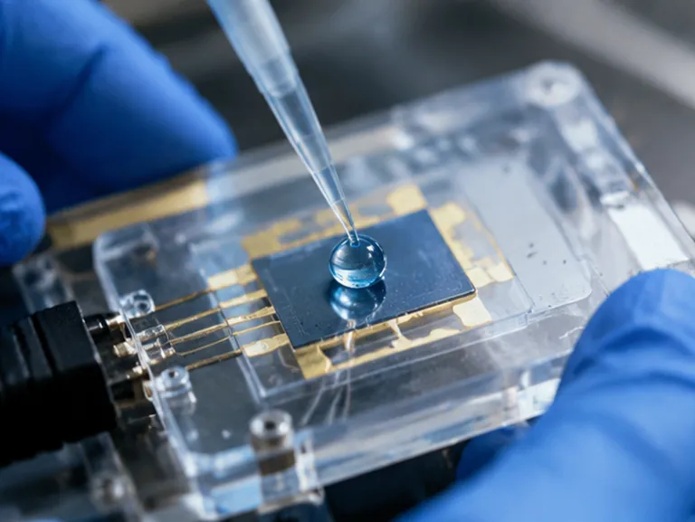
A screen-printed electrode provides a practical foundation for portable and scalable electrochemical immunosensors. Yet its performance depends on more than electrical conductivity.
The surface must support a controlled biological interface.
Developers need to manage:
- Active surface area.
- Coating density.
- Immobilization chemistry.
- Protein orientation.
- Blocking-layer uniformity.
- Electron-transfer properties.
- Resistance to nonspecific protein adsorption.
A high coating density can increase the amount of competitor available for antibody binding. But beyond a certain point, a crowded surface may create steric barriers or produce heterogeneous binding sites. The highest mass of immobilized protein is not automatically the highest analytical performance.
The useful target is an accessible, uniform, and reproducible competitor layer.
Blocking agents are equally important. Any unoccupied region of the electrode can attract proteins from the sample or assay mixture. These proteins may alter the electrochemical interface, trap labeled antibody, or create variable background current.
A blocking strategy must suppress nonspecific binding without masking the immobilized hapten or interfering with electron transfer.
Signal-to-Noise Ratio Is the Real Sensitivity Metric
The practical detection limit depends on whether the system can distinguish a target-driven signal change from baseline variation.
This is the signal-to-noise problem.
Signal can be increased through:
- A high-affinity antibody.
- An active HRP conjugate.
- An optimized substrate concentration.
- An efficient electrode interface.
- An appropriate incubation design.
Noise must be controlled through:
- Consistent electrode coating.
- Effective blocking.
- Low nonspecific antibody adsorption.
- Stable enzyme conjugates.
- Controlled washing.
- Matrix-compatible assay buffers.
- Tight temperature and timing control.
In a competitive assay, a small change near the upper signal range may determine whether a trace concentration is measurable. The developer must therefore characterize blank variability with the same seriousness given to analytical signal.
A claimed low detection limit is meaningful only when it remains stable across operators, reagent lots, electrode batches, and realistic sample matrices.
Matrix Effects Turn a Beautiful Assay into a Difficult Product
A sensor may reach an impressive detection limit in buffer and fail in whole blood.
Serum and other biological matrices contain abundant proteins, salts, lipids, endogenous antibodies, and chemically active compounds. These components can alter antibody binding, affect enzyme activity, change electrode wetting, and interfere with electron transfer.
Heterophilic antibodies and other nonspecific interactions are especially troublesome. They can create false signal, consume labeled antibody, or produce sample-dependent changes that do not reflect target concentration.
The solution is rarely a single additive. Matrix robustness usually requires coordinated optimization of:
- Sample dilution.
- Buffer composition.
- Ionic strength.
- Surfactants.
- Protein blockers.
- Non-immune serum components.
- Incubation time.
- Washing conditions.
- Surface chemistry.
This introduces a central trade-off. Increasing surface activity can improve signal generation, while increasing blocking strength can improve background control. The final formulation must preserve enough accessible binding capacity without allowing the sample matrix to dominate the measurement.
The Cost of Extreme Sensitivity
Every additional order of magnitude in sensitivity usually creates a new operational requirement.
A highly sensitive competitive electrochemical immunosensor may need:
- Precisely timed incubations.
- Controlled reagent ratios.
- Multiple washing steps.
- Careful substrate preparation.
- Strict temperature management.
- Calibrators that remain stable across the working range.
- Operators trained to avoid timing and pipetting variation.
Homogeneous formats may be easier to operate because they eliminate washing or separation. But convenience can come at the cost of lower sensitivity or weaker matrix tolerance.
The correct choice depends on the intended use.
| Development priority | Primary design emphasis |
|---|---|
| Lowest possible detection limit | Antibody affinity, slow dissociation, and catalytic amplification |
| Reproducible manufacturing | Characterized conjugates and validated lot consistency |
| Direct testing in complex samples | Blocking strategy, matrix compatibility, and interface control |
| Simple workflow | Reduced steps, stable reagents, and tolerance to timing variation |
| Portable or decentralized testing | Screen-printed electrodes, compact readout, and robust formulation |
Sensitivity is not free. It is purchased through control.
From Proof of Concept to Manufacturable Assay
The transition from a promising laboratory result to a commercial IVD product often exposes weaknesses that were invisible during early development.
A proof-of-concept experiment may use one antibody lot, one conjugate preparation, one electrode batch, and carefully prepared buffer. A production assay must survive the variation inherent in biological materials and manufacturing processes.
Critical controls include:
- Identity and purity of antibody materials.
- Affinity and specificity characterization.
- Hapten-to-carrier conjugation ratio.
- HRP activity after conjugation.
- Functional performance of each lot.
- Stability under intended storage conditions.
- Electrode coating uniformity.
- Calibration-curve shift over time.
- Matrix recovery and interference behavior.
Raw-material quality is therefore a strategic part of assay design. If the antibody affinity changes between lots, the calibration curve can move. If the hapten loading changes, the surface competition can change. If the HRP conjugate loses activity, the analytical signal can collapse even when the binding chemistry remains intact.
Reliable sourcing reduces the need to repeatedly re-optimize the entire system.
A Practical Development Sequence

A disciplined development program can reduce wasted optimization cycles.
1. Define the Measurement Context
Specify the target concentration range, sample type, intended turnaround time, instrument format, and acceptable workflow complexity.
2. Select and Characterize the Receptor
Screen monoclonal antibodies for affinity, kinetics, specificity, matrix tolerance, and stability.
3. Build the Competitive Architecture
Establish the relationship between free target, immobilized hapten-protein conjugate, and labeled antibody before optimizing the electrode in detail.
4. Optimize the Competitor
Control hapten orientation, loading, immobilization density, and lot consistency.
5. Develop the Enzyme Conjugate
Balance HRP activity with antibody functionality, steric accessibility, and storage stability.
6. Engineer the Interface
Optimize coating, blocking, washing, and electrochemical conditions to reduce nonspecific background.
7. Challenge the Assay with Real Matrices
Test recovery, interference, cross-reactivity, and precision using representative samples rather than buffer alone.
8. Establish Supply and Release Controls
Define material specifications and functional acceptance criteria before process scale-up.
This sequence reflects a simple truth: the electrode cannot compensate for an unsuitable receptor, and an excellent receptor cannot compensate for uncontrolled surface chemistry.
Choosing Raw Materials as a Business Decision
For diagnostic manufacturers, laboratories, and research institutes, raw-material selection affects more than technical performance.
It affects development speed, transfer risk, inventory planning, regulatory documentation, and the likelihood of maintaining a stable product after launch.
A supplier should be evaluated on more than catalog availability. Important questions include:
- Can the material be supplied consistently at development and production scale?
- Are analytical and functional specifications documented?
- Is lot-to-lot performance monitored?
- Can the supplier support conjugation and formulation decisions?
- Are technical services available when matrix effects appear?
- Can the material package support the transition from concept to clinic?
CamelBio provides diagnostic manufacturers, labs, and research institutes with one-stop access to IVD raw materials, technical services, and consulting across the development pathway.
That support can include high-affinity antibodies, hapten-carrier conjugates, HRP-labeled reagents, and technical guidance for addressing matrix effects, interface chemistry, and lot-to-lot variability.
Final Perspective: Sensitivity Is an Integrated Property

The most sensitive electrochemical immunosensor is not created by selecting the most conductive electrode or the most active enzyme in isolation.
It is created by aligning several constraints:
- The target's molecular geometry determines the assay architecture.
- The antibody establishes the thermodynamic and kinetic foundation.
- The hapten-protein conjugate controls competitive presentation.
- HRP provides catalytic amplification.
- The electrode interface determines how much of the theoretical signal survives in practice.
- Blocking and formulation determine whether the assay can function in real samples.
- Raw-material consistency determines whether the performance can be manufactured.
Small-molecule detection rewards precision because its margin for error is narrow. Every uncontrolled interaction becomes part of the background. Every shift in affinity or conjugation can move the calibration curve.
The engineering challenge is to make the entire system behave as one reproducible measurement rather than as a collection of individually impressive components.
For help selecting materials and integrating assay performance from concept to clinic, connect with Contact Our Experts.
Related Products
- Anti-MMP1 Rabbit Monoclonal Antibody for WB, ELISA - P03956
- Anti-SOCS7 Monoclonal Antibody for WB, ELISA - O14512
- Anti-SNRK Polyclonal Antibody for WB and ELISA - Q9NRH2
- Anti-IDH1 Rabbit Monoclonal Antibody for WB, IF/ICC, ELISA - O75874
- Anti-ABI3 Rabbit Monoclonal Antibody for WB, IHC-P, ELISA - Q9P2A4
![[KD Validated] Anti-TIMM10 Rabbit Monoclonal Antibody for WB, IHC-P, IF/ICC, ELISA - P62072](https://image.camelbiological.com/images/products/2969/main_image_6a633b15dcb875.18347145-small.webp)
![Anti-eIF4E Rabbit Monoclonal Antibody [KD Validated] for WB, IF/ICC, IP, ELISA - P06730](https://image.camelbiological.com/images/products/4940/main_image_6a636cf2795024.14314593-small.webp)