Polyclonal Antibodies
Anti-NRAS Rabbit Polyclonal Antibody for WB - P01111
Item Number : CM0028193
Price varies based on specs and customizations
- Application
- WB, IF/ICC, ELISA
- Cross Reactivity
- Human, Mouse, Rat
- Protein Weight
- 21kDa

Shipping:
Contact us to get shipping details Enjoy On-time Dispatch Guarantee.
Why Choose Us
Easy ordering process, quality products, and dedicated support for your business success.
Core Product Specifications and Parameters
| Parameter | Value |
|---|---|
| Product Name | NRAS Rabbit pAb |
| Remarks/Alias | NS6; CMNS; KRAS; NCMS; ALPS4; N-ras; NRAS1; NRAS |
| Species | Human |
| Gene ID (Human) | 4893 |
| Gene ID | 4893 |
| Immunogen | A synthetic peptide corresponding to a sequence within amino acids 90-189 of human NRAS (NP_002515.1). |
| Source | Rabbit |
| Category | Polyclonal Antibodies |
| Application | WB, IF/ICC, ELISA |
| Cross Reactivity | Human, Mouse, Rat |
| SWISS | P01111 |
| Protein Weight | 21kDa |
| Shipping | Ice bag |
Biological Background: NRAS Function and Localization
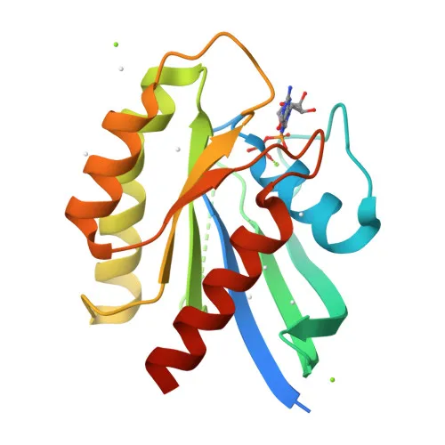
- NRAS is a member of the Ras small GTPase family, also known as Transforming protein N-Ras (gene synonym HRAS1), with aliases NS6, CMNS, KRAS, NCMS, ALPS4, N-ras, and NRAS1.
- It functions as a signal transducer in the Ras-MAPK signaling pathway, regulating cell proliferation and survival. Related references: PMID:30712867
- NRAS possesses intrinsic GTPase activity, cycling between an active GTP-bound and inactive GDP-bound state. Related references: PMID:30712867
- The protein is recognized by LZTR1, which mediates its ubiquitination by a BCR (BTB-CUL3-RBX1) E3 ubiquitin-protein ligase complex. Related references: PMID:40934300
- Subcellularly, NRAS is localized to the cell membrane and the Golgi apparatus membrane.
- Post-translational modifications include acetylation, methylation, palmitoylation, prenylation, phosphorylation, and isopeptide bond formation, as indicated by UniProt keywords.
- NRAS is classified as a proto-oncogene; mutations are associated with various cancers (keyword: Disease variant).
Experimental Guidance and Technical Tips
- For Western blot, NRAS is predicted at ~21 kDa; however, prenylation and membrane association may cause shifts. Validate the band size in your specific sample system.
- In immunofluorescence/immunocytochemistry, expect cell membrane and Golgi apparatus staining. Co-localization studies with compartment markers are recommended.
- The immunogen sequence (amino acids 90–189) is conserved across human, mouse, and rat, supporting cross-reactivity in these species; still, verify reactivity in your experimental model.
CamelBio: Your One-Stop Sourcing Bridge
CamelBio is your one-stop partner for IVD raw-material sourcing, offering validated antibodies like this NRAS polyclonal antibody, along with bulk ancillary reagents and custom rare-target monoclonal antibody development. Whether advancing oncology diagnostics or research, we bridge your supply needs from concept to clinic.
REQUEST A QUOTE
Our professional team will reply to you within one business day. Please feel free to contact us!
Related Articles
ROX in qPCR: The Quiet Reference That Makes Diagnostic Signals Trustworthy
Learn how ROX normalizes qPCR fluorescence, stabilizes Ct calls, and shapes the trade-off between instrument compatibility and multiplexing.
From Molecular Recognition to Reliable Results: Engineering Biosensors for Rapid Safety Testing
How antibodies and enzymes turn invisible molecular events into reliable biosensor signals, and how material quality determines test performance.
qPCR Primer Design: The Small Decisions That Determine Assay Reliability
A practical guide to qPCR primer design, from amplicon length and 3' stability to validation for efficient, reproducible diagnostics.
ROX in Real-Time PCR: The Optical Trade-Off Behind Precision and Multiplexing
Understand how ROX normalization corrects qPCR signal variation, how optics consume channels, and how to choose the right master mix.
From Fluorescence to Diagnostic Truth: Choosing Passive Reference Dyes for qPCR Master Mixes
Learn how passive reference dyes stabilize qPCR signals, improve diagnostic reproducibility, and guide ROX formulation across instrument platforms.
When a Surface Becomes a Sensor: Choosing Between QCM, SAW, and Six Classes of Quality Detection
Explore six sensor families, how QCM and SAW convert molecular binding into frequency shifts, and why surface chemistry determines diagnostic reliability.
The Silent Well: How No Template Controls Protect Molecular Diagnostic Integrity
A practical guide to No Template Controls: how unexpected qPCR signals reveal contamination, reagent impurities, artifacts, and broken workflow controls.
Why Minor Groove Binders Turn Short Hydrolysis Probes into Precision Diagnostic Tools
MGB chemistry improves probe stability, SNP discrimination, signal-to-noise ratio, and low-copy detection in demanding molecular assays.
Designing Generic Antibodies: How Hapten Engineering Enables Broad-Spectrum Pesticide Detection
Learn how strategic hapten design exposes conserved pesticide structures, improves antibody cross-reactivity, and guides reliable multi-residue assays.
The Signal You Lose Before the qPCR Run Begins: A Practical System for Protecting Primers and Fluorogenic Probes
Learn how aliquoting, dark storage, controlled thawing, and buffer choice protect qPCR fluorescence, Cq reproducibility, and assay sensitivity.
From a 36-Hour Wait to a 30-Minute Decision: The Workflow Advantage of Rapid Antibody-Based Immunosensors
Rapid antibody-based immunosensors compress analyte detection from 24–36 hours to about 30 minutes, changing workflows, decisions, and assay design.

Engineering High-Sensitivity Electrochemical Immunosensors for Small-Molecule Detection
How competitive assay design, high-affinity antibodies, HRP labels, and interface control enable reliable trace small-molecule detection.