The Weak Signal Usually Starts Before the PCR Run
A real-time PCR assay can fail quietly.
The instrument reports amplification, but the curve rises late. The endpoint fluorescence is modest. Replicates drift. A developer may first suspect the fluorophore, quencher, polymerase, or instrument calibration.
Often, the real problem was decided earlier, when the probe sequence and amplicon architecture were chosen.
A fluorogenic hydrolysis probe produces a signal only when several events occur in the correct order:
- The probe must find and bind its target.
- It must remain hybridized as the polymerase approaches.
- The polymerase must cleave the probe during extension.
- The reporter must be separated from the quencher.
- The released fluorescence must rise above background consistently.
This makes signal strength a kinetic problem, not merely a chemistry problem.
The probe must win a race against polymerase extension. Every cycle gives it one opportunity to be fully hybridized at precisely the moment the enzyme arrives.
What the Polymerase Is Actually Measuring
In a hydrolysis assay, the reporter and quencher begin in close proximity. The quencher suppresses the reporter’s fluorescence.
During extension, the 5′ to 3′ exonuclease activity of Taq polymerase degrades a probe hybridized to the target strand. Cleavage physically separates reporter from quencher, allowing fluorescence to accumulate.
The reaction therefore depends on timing.
If the probe is unbound, partially melted, folded into a hairpin, or located too far from the primer, the polymerase may pass without producing an efficient cleavage event. The assay may still amplify, but fewer probes are cleaved per cycle.
That difference appears as:
- Delayed Cq values
- Shallow amplification curves
- Lower endpoint fluorescence
- Reduced signal-to-noise ratio
- Greater sensitivity to small changes in sample or reagent concentration
A useful mental model is simple:
Signal is strongest when probe hybridization is complete before polymerase extension begins, and cleavage follows almost immediately after extension starts.
Four Design Decisions Control the Signal
1. Set Probe Tm Above the Primer Annealing Temperature
A practical target for most hydrolysis probes is a melting temperature of 68–70°C.
This is typically 8–10°C above a primer annealing temperature of approximately 58–60°C. The offset gives the probe a thermodynamic advantage. At the annealing and extension phase, the probe should remain stably bound while the primers support efficient amplification.
If the probe Tm is too low, it may dissociate before polymerase reaches it. The enzyme cannot efficiently cleave a probe that is not correctly duplexed with the target.
Use the same salt-adjusted thermodynamic model for both primers and probes. A nearest-neighbor model that accounts for sodium and magnesium concentrations is preferable to comparing values generated by unrelated calculators.
The important variable is not the absolute number alone. It is the relationship between probe and primer Tm.
| Parameter | Practical target | Reason |
|---|---|---|
| Probe Tm | 68–70°C | Maintains stable target binding during extension |
| Primer Tm | Approximately 58–60°C | Supports controlled annealing |
| Probe-primer Tm difference | 8–10°C | Gives the probe a hybridization advantage |
| Excessively high probe Tm | Above approximately 72°C | May reduce mismatch discrimination |
A higher Tm is not automatically better.
When probe Tm becomes excessive, the probe may tolerate mismatches that should have prevented binding. In a diagnostic assay, this can weaken discrimination between a target and a closely related non-target.
The goal is not maximum binding at any cost. The goal is stable, specific binding under the actual cycling conditions.
2. Put the Primer and Probe in Close Physical Proximity
The polymerase begins extending from the upstream primer. The closer the probe is to the primer’s 3′ end, the sooner the enzyme encounters it.
A practical design range is 0–5 bases between the primer’s 3′ end and the probe’s 5′ end.
This spacing reduces the time available for the probe to dissociate and shortens the interval between extension and cleavage. It also commonly supports a compact amplicon of approximately 50–100 bp.
The effect is easy to underestimate because the difference may be only a few nucleotides. But PCR is repeated thousands or millions of times. A small kinetic delay is repeated in every cycle and can become a visible loss of signal.
| Primer-probe spacing | Expected effect |
|---|---|
| 0–2 bases | Very rapid polymerase encounter and cleavage |
| 3–5 bases | Usually compatible with strong signal kinetics |
| More than 5 bases | Increased extension distance and possible kinetic delay |
| Very long spacing | Greater dependence on probe stability and reaction conditions |
Close spacing should still be balanced with sequence quality. A primer should not be forced into a region with strong secondary structure, poor specificity, or unfavorable 3′ composition simply to reduce distance.
The best design is the shortest reliable path from primer extension to probe cleavage.
3. Choose a Target Region That Supports Fast, Stable Hybridization
Sequence composition affects both thermodynamics and fluorescence quality.
A target region with strong cytosine-related binding potential can increase duplex stability because C-G base pairs form three hydrogen bonds. However, the sequence must be evaluated in the correct orientation.
A C-rich target may require a probe with elevated guanine content. That creates a second design risk at the reporter-bearing end.
Protect the 5′ Reporter End
Avoid more than two consecutive guanines near the probe’s 5′ end.
Guanine can reduce reporter fluorescence through photoinduced electron transfer, even when the quencher remains chemically intact. The result is lower baseline performance and a weaker apparent signal.
The sequence may therefore bind well while still producing poor fluorescence.
Check for Secondary Structure
A probe that folds into a stable hairpin or self-dimer is temporarily unavailable for target binding.
Evaluate:
- Probe hairpins
- Probe self-dimers
- Primer-dimers
- Cross-dimers between primers and probe
- Long homopolymer runs
- Excessive G-rich stretches
- Potential G-quadruplex-forming regions
If a target is highly G-rich, consider adjusting probe length or using modified bases such as Locked Nucleic Acid where appropriate. Modifications can raise Tm without requiring an excessive increase in guanine content.
The sequence should be treated as a physical object with structure, not just as a string of letters.
4. Validate the Primers Before Blaming the Probe
A probe cannot compensate for inefficient amplification.
If the primers generate dimers, nonspecific products, or uneven amplification, they consume polymerase, nucleotides, and template before the probe can generate a clean signal.
Primer validation should include:
- A single, sharp melt-curve product when using SYBR Green I
- Amplification efficiency between 90% and 110%
- A standard-curve slope approximately between -3.6 and -3.1
- Absence of meaningful primer-dimer formation
- Sequence confirmation of the intended amplicon
- Exact verification of the probe-binding site
The final check matters because a single nucleotide mismatch can substantially weaken probe hybridization. In a research experiment, that may appear as variability. In a diagnostic workflow, it can become a false negative or a loss of confidence in the result.
The Design Is a System, Not a Collection of Good Numbers
A probe with a Tm of 70°C may still perform poorly if it forms a hairpin.
A probe positioned two bases from the primer may still produce weak signal if the primers amplify inefficiently.
A C-rich binding site may still be unsuitable if the resulting probe contains a guanine cluster beside the reporter.
The relevant design unit is the complete assay architecture.
| Design criterion | Recommended specification | Kinetic or analytical benefit |
|---|---|---|
| Probe Tm | 68–70°C | Promotes complete hybridization before extension |
| Tm offset | 8–10°C above primer annealing temperature | Gives the probe a stability advantage |
| Primer-probe spacing | 0–5 bases | Enables rapid cleavage after extension begins |
| Amplicon length | Commonly 50–100 bp | Supports efficient amplification and low kinetic delay |
| Target composition | Favor stable hybridization without excessive G-richness | Improves binding while protecting fluorescence |
| 5′ guanine content | No more than two consecutive G residues near reporter | Reduces reporter quenching |
| Primer efficiency | 90–110% | Confirms that amplification is not consuming assay capacity inefficiently |
| Product specificity | One intended amplicon | Prevents nonspecific products from diluting signal |
Where Optimization Can Go Wrong
Pushing Tm Too High
A very stable probe can appear attractive, especially when the initial assay has a weak curve.
But excessive stability can reduce mismatch discrimination. This is dangerous when the assay must distinguish:
- A single-nucleotide variant
- A pathogen from a close relative
- A mutant allele from wild type
- A target sequence from a homologous background
Stay near the 68–70°C range unless the assay has a specific reason to require a different design. Use probe length and carefully controlled composition before resorting to extreme GC content.
Making the Amplicon Too Short
Short amplicons generally support rapid amplification, but there is a lower practical limit.
Below approximately 50 bp, primer placement becomes constrained. The sequence may also become more sensitive to local secondary structure or binding-site interference. A design that looks efficient on paper may become fragile across sample types.
A range of 60–100 bp is often a robust compromise for general qPCR assay development.
Treating C-Richness as an Absolute Rule
Stable binding is valuable, but composition must be balanced.
A very C-rich target can produce a G-rich probe. Excessive guanine may encourage aggregation, G-quadruplex formation, or reporter quenching. The strongest theoretical duplex is not always the strongest practical fluorescent probe.
Sequence quality should be judged across four dimensions:
- Hybridization stability
- Specificity
- Structural behavior
- Reporter compatibility
Match the Design to the Assay’s Real Priority
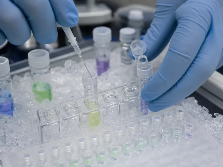
There is no single optimal probe design for every application.
For Low-Copy Detection
Prioritize:
- Primer-probe spacing of 0–5 bases
- Probe Tm around 69–70°C
- Primer efficiency above 95%
- Minimal secondary structure
- No guanine cluster near the 5′ reporter
The objective is to maximize the number of successful cleavage events from limited starting material.
For Multiplex Assays
Prioritize:
- Amplicons around 60–80 bp
- Consistent probe Tm across targets
- Comparable primer efficiencies
- Balanced sequence composition
- Similar expected cleavage kinetics
In a multiplex reaction, one highly efficient target can consume shared reagents faster than another. Harmonized kinetics help prevent one assay from dominating the reaction.
For SNP or Variant Discrimination
Prioritize:
- A discriminatory base near the center of the probe
- Probe Tm near 68°C
- Carefully controlled mismatch sensitivity
- Sufficient amplicon flexibility for specific primer placement
A slightly longer amplicon can be worthwhile if it allows the primers and probe to avoid nonspecific or structurally difficult regions.
For Rapid Time-to-Result
Prioritize:
- An amplicon of approximately 50–70 bp
- A probe Tm near 70°C
- A combined annealing-extension step near 60°C, where validated
- Efficient primer design with minimal side reactions
Shorter extension paths and stable probes can produce steeper curves and earlier Cq values, provided specificity is preserved.
A Practical Development Sequence
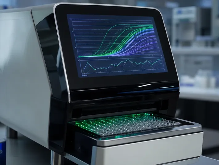
A disciplined workflow reduces the temptation to solve every problem by changing the probe.
Step 1: Define the Analytical Requirement
Clarify whether the assay is optimized for sensitivity, specificity, multiplex balance, variant discrimination, or speed.
That decision determines which trade-offs are acceptable.
Step 2: Select the Amplicon
Search for a target region that supports:
- A reliable 50–100 bp amplicon
- Close primer-probe positioning
- Low secondary structure
- A unique sequence
- An accurately confirmed probe-binding site
Step 3: Design the Primers
Calculate primer Tm using the same thermodynamic assumptions that will be used for the probe. Screen for dimers, hairpins, and nonspecific binding.
Step 4: Design the Probe
Aim for 68–70°C. Check the 5′ reporter environment carefully. Avoid excessive guanine, especially consecutive G residues, while preserving target specificity.
Step 5: Validate the Amplification System
Use a standard curve and melt-curve analysis where appropriate. Confirm efficiency, product identity, and the absence of competing products.
Step 6: Investigate Weak Signal Systematically
When the curve is weak, review the following in order:
| Question | Failure it may reveal |
|---|---|
| Is the probe fully hybridized at the extension temperature? | Low probe Tm or mismatch |
| Is the primer close enough to the probe? | Delayed cleavage |
| Does the probe form a hairpin or dimer? | Poor target availability |
| Is the 5′ end affected by guanine quenching? | Low fluorescence despite binding |
| Are the primers efficient and specific? | Reagent loss to side reactions |
| Does the amplicon contain the intended sequence? | Template mismatch or design error |
This sequence keeps troubleshooting anchored to mechanism.
From Sequence Design to Reliable Diagnostic Production
In an academic experiment, a weak signal may lead to another round of oligo design.
In a diagnostic development program, the consequences are larger. A design revision can affect raw material purchasing, lot qualification, analytical validation, documentation, transfer, and eventual clinical performance.
That is why probe design should be connected to the rest of the assay development workflow.
CamelBio supports diagnostic manufacturers, laboratories, and research institutes with one-stop access to IVD raw materials, technical services, and consulting. Its support can extend from early assay concept and oligo requirements through optimization and the transition toward clinical use.
The value is not limited to obtaining a probe sequence. It lies in coordinating the materials and technical decisions that determine whether the sequence performs reliably in the complete reaction.
Strong fluorescence is built cycle by cycle.
It begins with a probe that binds early, remains present, and is cleaved at the right moment. To connect probe design with dependable materials and technical support from concept to clinic, speak with Contact Our Experts.
Related Products
- Monoclonal Mouse IgG1 Isotype Control Antibody (Conjugate) for Flow Cytometry - Mouse IgG 1 isotype control
- Anti-BOLL Polyclonal Antibody for WB, ELISA - Q8N9W6
- Anti-ABI3 Rabbit Monoclonal Antibody for WB, IHC-P, ELISA - Q9P2A4
- Anti-pro Caspase-1 + p10 + p12 Rabbit mAb for WB, IP, ELISA - P29466
- Anti-Olig2 Rabbit Monoclonal Antibody for WB, ELISA - Q13516
Related Articles
- Why Direct Hapten Coupling Changes the Economics of Small-Molecule Biosensors
- The Silent Well: How No Template Controls Protect Molecular Diagnostic Integrity
- Two Positive Controls, One Clear Answer: Isolating RT and PCR Failure in qRT-PCR Diagnostics
- When the Dye Sees Everything: A Systematic Strategy for Cleaner, More Reliable qPCR
- From Molecular Recognition to Reliable Results: Engineering Biosensors for Rapid Safety Testing