Polyclonal Antibodies
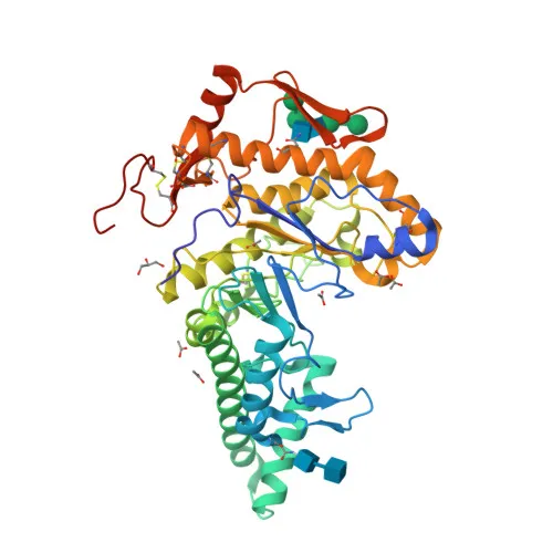
Anti-HYAL1 Rabbit Polyclonal Antibody for WB, IHC-P, IF/ICC, ELISA - Q12794
Item Number : CM0027167
Price varies based on specs and customizations
- Application
- WB, IHC-P, IF/ICC, ELISA
- Cross Reactivity
- Human, Mouse, Rat
- Protein Weight
- 48kDa

Shipping:
Contact us to get shipping details Enjoy On-time Dispatch Guarantee.
Why Choose Us
Easy ordering process, quality products, and dedicated support for your business success.
Core Product Specifications and Parameters
| Parameter | Value |
|---|---|
| Product Name | HYAL1 Rabbit pAb |
| Remarks/Alias | MPS9; NAT6; LUCA1; HYAL-1; HYAL1 |
| Species | Human |
| Gene ID (Human) | 3373 |
| Gene ID | 3373 |
| Immunogen | Recombinant protein |
| Source | Rabbit |
| Category | Polyclonal Antibodies |
| Application | WB, IHC-P, IF/ICC, ELISA |
| Cross Reactivity | Human, Mouse, Rat |
| SWISS | Q12794 |
| Protein Weight | 48kDa |
| Shipping | Ice bag |
Biological Background: HYAL1 Function and Localization
- HYAL1 (Hyaluronidase-1) is a lysosomal and secreted hydrolase that degrades hyaluronic acid, a major component of the extracellular matrix.
- It belongs to the hyaluronidase family and contains an EGF-like domain and disulfide bonds critical for its structure and function.
- HYAL1 is highly expressed in liver, kidney, and heart, with lower expression in lung, placenta, and skeletal muscle; it is absent in adult brain.
- Multiple isoforms exist due to alternative splicing; for example, isoform 1 is restricted to bladder and prostate cancer cells, while isoforms 3-6 are found in normal and tumor bladder tissues.
- The protein may promote tumor progression by degrading extracellular matrix hyaluronan, facilitating invasion and metastasis.
- It may also modulate cell growth by blocking TGFB1-enhanced proliferation.
- Subcellularly, HYAL1 localizes to lysosomes and is also secreted into the extracellular milieu.
- Genetic variants in HYAL1 are associated with mucopolysaccharidosis type IX, a lysosomal storage disorder.
Experimental Guidance and Technical Tips
- For Western blot (WB), the predicted molecular weight is 48 kDa; consider validating the detected band using appropriate positive controls and lysates from HYAL1-expressing tissues (e.g., liver, kidney).
- For immunohistochemistry (IHC-P) and immunofluorescence (IF/ICC), antigen retrieval conditions may need optimization. Test both enzymatic and heat-mediated methods on your specific tissue or cell samples.
- The antibody shows cross-reactivity with mouse and rat; we recommend verifying specificity and sensitivity in the relevant species model before large-scale experiments.
- Given the polyclonal nature and the immunogen spanning a large region (aa 136-435), it may recognize multiple HYAL1 isoforms; confirm band patterns against known isoform profiles.
CamelBio: Your One-Stop Sourcing Bridge
CamelBio streamlines IVD raw-material procurement for diagnostic developers and research labs. This anti-HYAL1 rabbit polyclonal antibody supports cancer biology and lysosomal storage disorder studies. Beyond individual antibodies, we offer validated antibody pairs, bulk ancillary reagents, and customized sourcing for rare-target proteins, helping you accelerate assay development from concept to clinic.
Product Datasheet
Anti-HYAL1 Rabbit Polyclonal Antibody for WB, IHC-P, IF/ICC, ELISA - Q12794
REQUEST A QUOTE
Our professional team will reply to you within one business day. Please feel free to contact us!