The Instrument Is Part of the Assay
A multiplex real-time PCR assay can fail before the first primer is synthesized.
The failure may begin with an apparently reasonable decision: choosing a bright fluorophore, adding another target, or transferring an established probe set to a different qPCR platform.
Then the amplification curves become unstable.
One channel rises when another target is abundant. A low-copy signal disappears into the baseline. Replicates disagree. The assay appears to have a chemistry problem, although the underlying constraint is optical.
The instrument is not a neutral container for the assay. Its light source, filters, detector, and normalization method define what the assay can reliably see.
For diagnostic manufacturers, this has an important consequence:
Fluorophore selection should begin with the instrument's optical architecture, not with a dye catalogue.
The optical system is the assay blueprint.
Why Brightness Alone Is a Weak Design Principle
A fluorophore is often judged by its brightness, extinction coefficient, or popularity across existing assays.
Those properties matter. They are not enough.
The instrument must first excite the dye efficiently. The emitted light must then pass through the correct filter and reach the detector with sufficient separation from background and neighboring channels.
A bright dye in the wrong optical window can perform worse than a less intense dye that matches the platform precisely.
The practical question is not:
Which fluorophore produces the strongest signal?
It is:
Which reporter creates the clearest, most stable spectral fingerprint within this instrument?
That fingerprint depends on four linked variables:
- Excitation wavelength and intensity
- Emission peak and bandwidth
- Filter configuration
- Detector sensitivity and readout timing
Changing one variable can alter the behavior of the entire multiplex panel.
Start with the Light Source
The light source determines which wavelengths are available to excite the reporters and how consistently that excitation is delivered over time.
Broad-Spectrum Lamps
Quartz tungsten-halogen and xenon lamps provide wide excitation coverage.
This flexibility allows instrument designers to define multiple channels with different filter combinations. It is useful when an assay requires a custom fluorophore panel or must support a broad selection of commercially available dyes.
The cost of that flexibility is operational complexity.
Lamps can generate heat, require warm-up, and degrade with use. Changes in output can affect calibration and long-term quantitative consistency. A platform may support several fluorophores in principle, while still requiring careful maintenance to preserve its original performance.
Broad-spectrum systems are often appropriate when:
- The assay portfolio is diverse
- Fluorophore flexibility is more important than hardware simplicity
- The platform must support multiple filter configurations
- The development team can manage calibration and lamp replacement
LED Arrays
LED arrays provide narrower and more stable excitation.
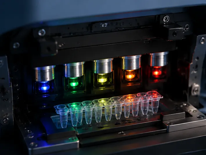
A multi-color LED system may offer four, five, or six predefined channels, sometimes including a channel designed for FRET-based detection. LEDs produce little heat, have long operating lifetimes, and offer predictable output for routine diagnostic workflows.
Their constraint is equally clear: the assay must fit the available wavelengths.
An LED-based platform can be highly reliable when its channel map is known early in development. It is less forgiving when a team attempts to add an unconventional fluorophore late in the design process.
LED systems are particularly attractive when:
- The assay uses a standardized fluorophore set
- High throughput and low maintenance are priorities
- The instrument will operate continuously in a clinical laboratory
- Reproducible excitation is more important than broad optical flexibility
Lasers
Lasers deliver intense, monochromatic excitation.
That intensity can improve detection of weak signals, especially when the optical path and detector are designed for it. But more excitation is not always better for a quantitative assay.
Repeated high-intensity exposure can accelerate photobleaching. As the reporter degrades, the amplification curve may no longer reflect the underlying accumulation of amplicon. A system optimized for maximum photon flux can therefore create a new source of quantitative error.
For low-copy targets, the better strategy is usually balanced:
- Use a bright, photostable reporter
- Minimize unnecessary excitation
- Control exposure time and intensity
- Characterize signal stability across the full cycling program
Sensitivity is not simply the ability to collect more photons. It is the ability to collect informative photons consistently.
The Detector Changes the Meaning of a Measurement
Two instruments can use the same fluorophore and produce different assay behavior because they collect the signal differently.
CCD Cameras: Simultaneous Imaging
A CCD camera can image many or all wells at the same point in the thermal cycle.
This creates strong temporal consistency. Every well is observed under nearly identical cycle timing, which is valuable when comparing amplification kinetics across a plate.
The optical path, however, can vary slightly from well to well. Differences in volume, positioning, plastic, or illumination may influence the measured intensity.
That is why some CCD-based systems use a passive reference dye such as ROX. The reference signal helps normalize optical variation.
This creates a design trade-off:
- Normalization can improve well-to-well consistency
- The passive dye occupies optical and chemical capacity
- A channel used for ROX may no longer be available for a target
- The master mix must be compatible with the reference dye requirement
ROX is therefore not a universal ingredient. It is a consequence of a particular measurement architecture.
PMT Scanners: Sequential, Sensitive Reading
Photomultiplier tubes can detect very low levels of light with high sensitivity.
In many systems, wells are measured sequentially rather than simultaneously. This can reduce background and improve low-copy detection, but it introduces a small timing difference between wells. A scan may take several seconds, and the plate is not observed at exactly one instant.
For routine endpoint interpretation, that time difference may be manageable. For assays that depend heavily on precise kinetic comparison, it must be evaluated during validation.
PMT-based systems are often suited to:
- Low-copy targets
- Low-background detection
- High analytical sensitivity
- Assays where photon-counting performance outweighs simultaneous acquisition
The central question is not whether a PMT is sensitive. It is whether the complete workflow accounts for sequential acquisition.
Filtered Photodiodes: Dedicated and Predictable
Filtered photodiodes are commonly paired with LED excitation.
They read targeted optical channels with relatively simple hardware and low maintenance requirements. This makes them practical for low- to mid-plex assays in which each channel has a defined role.
Their advantage is predictability. Their limitation is flexibility.
A photodiode platform can be an efficient choice when the assay panel is stable, the number of targets is controlled, and manufacturing cost and serviceability matter as much as maximum sensitivity.
Channel Count Is Not Multiplex Capacity
An instrument with six optical channels does not automatically support a six-plex diagnostic assay.
The true limit is spectral separation.
Every reporter emits across a range of wavelengths. If the emission profile of one dye extends into a neighboring filter band, the detector records a mixture rather than a clean target-specific signal.
This is spectral crosstalk.
Crosstalk becomes especially dangerous when one target is present at a high concentration and another is near its limit of detection. The strong channel can raise the apparent baseline of the weak channel, producing false positives, distorted quantification, or inconsistent threshold cycles.
A useful way to assess a multiplex panel is to ask:
| Design question | Why it matters |
|---|---|
| Where is each reporter's emission peak? | The peak must align with the instrument's detection window. |
| How wide is each emission spectrum? | Broad emission increases the risk of spillover. |
| How narrow are the instrument filters? | Narrow filters improve separation but can reduce collected signal. |
| Can the software compensate for spectral overlap? | Compensation helps, but cannot recover information that the optics never separated. |
| What happens when one target is highly abundant? | The strongest channel often exposes weaknesses in the panel. |
The number printed in the instrument brochure is only the starting point.
Crosstalk, not channel count, is the real multiplex limit.
Build a Spectral Map Before Ordering Probes
A robust assay design begins with a channel map.
For each candidate reporter, record:
- Excitation maximum
- Emission maximum
- Relative brightness
- Photostability
- Compatibility with the instrument filters
- Expected spillover into adjacent channels
- Compatibility with the selected quencher
- Availability across intended platforms
Then evaluate the panel as a system.
A fluorophore that performs well in a singleplex assay may become unsuitable when paired with another dye. Likewise, a panel that works on one instrument may lose separation on another because the filter bandwidths and detector response curves are different.
The transfer question should therefore be specific:
Does this platform preserve the spectral distance and signal-to-background ratio that the original validation depended on?
“Compatible with qPCR” is too broad a category to answer that question.
Probe Chemistry Completes the Optical Design
The optical block cannot compensate for a noisy probe.
In a fluorogenic probe, the reporter and quencher are designed to remain optically quiet until amplification separates or changes their relationship. Hydrolysis probes use enzymatic cleavage to release the reporter from the quencher. Molecular beacons use a conformational change.
Both designs depend on chemical quality.
A poorly purified dual-labeled oligonucleotide can contribute residual fluorescence to the baseline. An unsuitable quencher may fail to absorb the reporter's emission efficiently. A fluorescent quencher may also introduce secondary signals into an already crowded panel.
For multiplex assays, the quencher should be selected with the full reporter set in mind.
Important considerations include:
- Broad absorbance across the reporter wavelengths
- Low residual fluorescence
- Minimal re-emission within active detection channels
- Consistent performance across probe lengths and sequences
- Chemical purity suitable for low-background detection
The objective is not merely to create a bright positive signal. It is to create a dark and stable negative state.
That negative state determines how confidently the system can recognize a weak positive.
The Background Has a Psychology of Its Own
A low-copy target does not fail dramatically. It disappears gradually.
A baseline rises by a small amount. A replicate crosses the threshold late. An operator sees an ambiguous curve and begins to question the sample, the extraction, or the reagent lot.
This is where background becomes more than an engineering specification. It changes human judgment.
High background encourages conservative thresholds. Excessive compensation encourages false confidence. Variable baselines make borderline results difficult to interpret and harder to defend during verification.
Two background sources deserve particular attention:
Autofluorescence
Plastic consumables, optical components, reagents, and sample matrices can emit light under excitation.
Low-fluorescence plastics and carefully selected consumables reduce this contribution. The effect should be characterized with the actual plate, master mix, and specimen matrix rather than with water alone.
Detector Dark Current
Photodetectors can generate a signal even without meaningful fluorescence.
Dark current becomes important when the assay is working near its detection limit. Detector cooling, active area, integration time, and instrument electronics all influence the noise floor.
The practical lesson is simple:
Limit-of-detection performance belongs to the complete optical and chemical system, not to the reporter molecule in isolation.
Multiplex Design Is a Trade-Off Between Three Clocks
Every real-time PCR assay operates against three forms of time:
- The reaction's amplification kinetics
- The instrument's acquisition timing
- The reporter's photochemical stability
A CCD system aligns the acquisition clock across wells but may require passive normalization. A PMT scanner provides strong low-light performance but introduces sequential timing. A high-power light source increases instantaneous signal but may shorten the reporter's useful lifetime.
These trade-offs can be summarized as follows:
| Priority | Optical direction | Main benefit | Main risk |
|---|---|---|---|
| High-throughput multiplexing | Multi-channel LEDs with narrow filters | Stable, repeatable channel excitation | Limited to predefined wavelengths |
| Low-copy sensitivity | PMT or high-sensitivity hybrid detection | Strong photon-counting performance | Sequential readout and timing skew |
| Operational simplicity | LEDs with filtered photodiodes | Long life and low maintenance | Lower flexibility for expanded panels |
| Cross-platform portability | Widely supported dyes such as FAM, HEX, and ROX | Easier transfer between instruments | Platform-specific normalization and filter differences |
| Maximum optical flexibility | Broad-spectrum lamp with configurable filters | Supports diverse fluorophore sets | Heat, calibration, and lamp aging |
| Photostability | Moderate controlled excitation | More stable quantitative curves | May require brighter or more efficient chemistry |
There is no universally superior optical architecture.
There is only a better match between architecture, diagnostic objective, and validation strategy.
A Practical Workflow for IVD Development
1. Freeze the Instrument Assumption Early
Define the intended instrument family before finalizing the fluorophore panel.
Record the available channels, excitation sources, emission filters, detector type, scan sequence, normalization requirements, and software compensation model.
2. Assign the Hardest Target First
The most difficult target is usually the low-copy, low-abundance, or clinically decisive target.
Give it the cleanest available channel and the most favorable signal-to-background conditions. Do not assign channels solely by convenience or by the order in which targets were added.
3. Stress-Test the Strongest Signal
Evaluate the panel with the expected maximum concentration of each target.
The strongest target should not create a false rise in neighboring channels. Test this using single-color controls, mixed high-positive controls, and clinically realistic sample backgrounds.
4. Validate the Chemistry and Optics Together
A probe's performance depends on its reporter, quencher, sequence, purification, master mix, and optical channel.
Changing any one of these can alter baseline fluorescence and apparent efficiency. Qualification should therefore use the final probe chemistry on the final instrument configuration.
5. Plan for Portability as a Separate Requirement
An assay intended for multiple platforms needs more than familiar dyes.
For every target, document:
- The instrument channel used
- Required passive reference dyes
- Spectral compensation settings
- Acceptable baseline behavior
- Minimum signal-to-background ratio
- Platform-specific limits of detection
- Any changes to the master mix
Portability is an engineering deliverable. It should be designed and verified, not assumed.
Choosing the Right Development Strategy
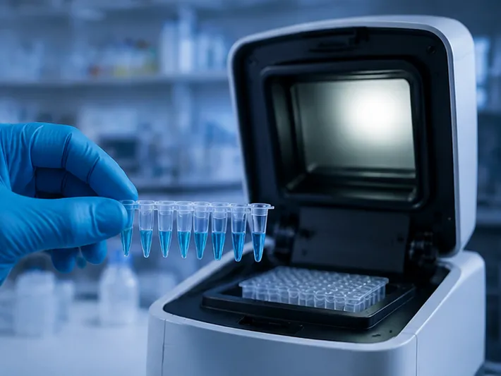
The best optical strategy depends on what the diagnostic product must accomplish.
For High-Plex Panels
Choose a platform with multiple discrete channels and narrow-band filters.
Prioritize reporters with separated emission profiles, then verify spillover under extreme target imbalance. Use broad-spectrum quenchers and include spectral calibration controls where necessary.
For Low-Copy Detection
Prioritize detector sensitivity and background control.
PMT-based or hybrid systems may offer an advantage, but the team must characterize sequential scan timing and photobleaching over the complete run. Bright, stable reporters are generally more useful than simply increasing excitation intensity.
For Distributed Laboratory Workflows
Favor stable, low-maintenance LED systems with predictable channel definitions.
A constrained dye set can be an advantage when it simplifies training, procurement, service, and quality control across multiple sites.
For Manufacturers Serving Multiple Platforms
Begin with broadly supported reporters, but validate each platform's actual filter set.
FAM, HEX, and ROX may provide a practical starting point, yet their behavior still depends on detector response, filter bandwidth, reference dye requirements, and software compensation.
The Engineering Decision That Saves the Most Time
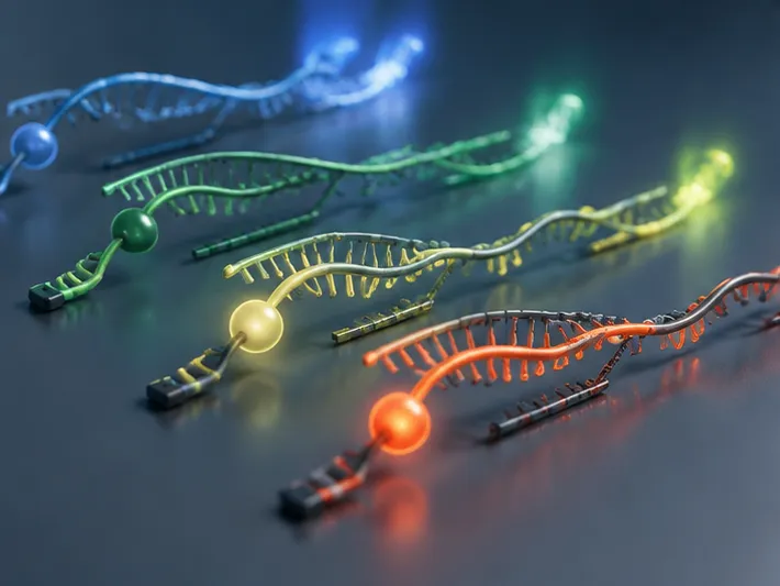
Many teams ask whether a new fluorophore can be added after the assay has already been designed.
A better question is whether the optical architecture can resolve the additional information without making interpretation less reliable.
Every added channel introduces more than one new dye. It adds:
- Another emission spectrum
- Another source of background
- Another compensation relationship
- Another probe and quencher interaction
- Another validation burden
- Another possible failure mode during platform transfer
Multiplexing is valuable because it increases information per reaction.
It is dangerous when it increases ambiguity faster than it increases information.
The strongest assay is not the one with the most colors. It is the one in which every color remains interpretable under the worst clinically plausible conditions.
From Optical Constraint to Diagnostic Confidence
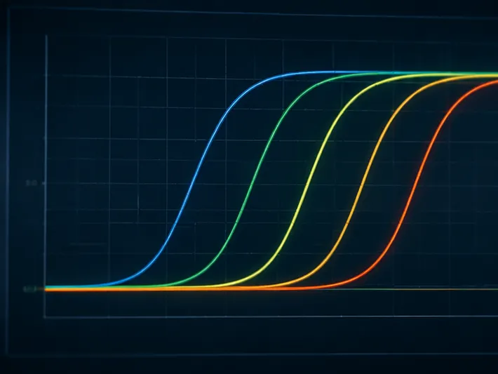
The optical system defines the boundaries.
Within those boundaries, assay developers still have meaningful choices: reporter selection, quencher chemistry, probe purification, excitation intensity, normalization strategy, and validation design.
The disciplined approach is to make those choices in the correct order:
- Understand the instrument architecture
- Map the usable spectral channels
- Assign reporters according to risk and target abundance
- Optimize probe and quencher chemistry
- Measure crosstalk and background under stress
- Validate performance across the intended platform set
This turns hardware limitations into a development roadmap.
CamelBio supports diagnostic manufacturers, laboratories, and research institutes with IVD raw materials, technical services, and consulting across the path from concept to clinic. When optical constraints are shaping your multiplex assay, start the design discussion with Contact Our Experts.
Related Products
- Anti-p63 Rabbit Monoclonal Antibody for WB, ELISA, ChIP - Q9H3D4
- Anti-STX1A Polyclonal Antibody for WB, IF/ICC, ELISA - Q16623
- Anti-CMIP Polyclonal Antibody for WB, IHC-P, ELISA - Q8IY22
- MonoMethyl-Histone H3-K79 Rabbit Polyclonal Antibody (Q16695 / P68431) for WB, ELISA, ChIP
- Anti-Tau Polyclonal Antibody for WB, IHC-P, IF/ICC, ELISA - P10636
Related Articles
- Where Ct Values Become Trustworthy: Establishing the qPCR Fluorescence Threshold
- qPCR Efficiency: The Mathematical Foundation of Reliable Molecular Diagnostics
- Designing Generic Antibodies: How Hapten Engineering Enables Broad-Spectrum Pesticide Detection
- When qPCR Fluorescence Becomes Data: The Engineering Logic of FAM, FRET, and Reliable Probe-Based Assays