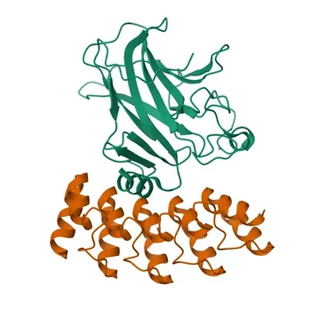
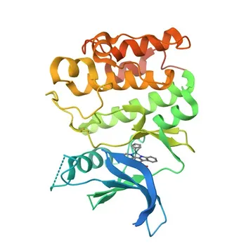
The two non-negotiable variables you must optimize are fusion-tag geometry and the donor-to-acceptor DNA ratio. When building a cell‑based protein‑protein interaction (PPI) assay for a technical service, the molecular design must confirm that the donor and acceptor tags do not block the interaction, and the transfection conditions must be titrated to deliver the highest possible signal window without distorting biology.
In technical‑service development, reproducible PPI data hinges on systematically testing both N‑ and C‑terminal fusions for each partner, then holding the donor plasmid constant while only the acceptor plasmid amount is varied. This locked‑donor transfection titration reveals the optimal dynamic range and assay sensitivity, turning a simple reporter assay into a robust, commercial‑grade service offering.
Molecular Parameters: Engineering the Perfect Reporter System
Why Fusion‑Tag Geometry Is the First Critical Decision
Any genetically encoded proximity assay—whether BRET, FRET, or split‑enzyme complementation—relies on fusing a reporter moiety to a protein of interest. A single fusion position can bury the binding interface or force the reporter out of the energy‑transfer radius.
You cannot guess which terminus will work. The donor (e.g., luciferase) and acceptor (e.g., fluorescent protein) must be tested at both the N‑Terminus and C‑Terminus of both candidate proteins. This creates a matrix of combinations that must be screened for biological activity and maximum energy transfer.
The BRET Benchmark: Matching Donor, Acceptor, and Geometry
In a classic BRET system, Renilla luciferase (~35 kDa) is the donor, emitting broad light peaking near 480 nm. An acceptor like EYFP (excitation 513 nm) is paired to create a 50‑Å Förster radius ($R_0$).
When the donor and acceptor are fused to interacting proteins, resonance energy transfer occurs only if the tags are positioned within this critical distance. An N‑terminal RLUC fusion may bring the donor too far from the acceptor’s binding‑induced location, while a C‑terminal fusion aligns them perfectly. Only empirical screening of all geometries guarantees that you preserve native interaction kinetics.
Transfection Parameters: The Locked‑Donor Titration Strategy
Why a Fixed Donor, Variable Acceptor Transfection is Mandatory
PPI assays report an interaction as a ratio of acceptor signal to donor signal. If the donor concentration drifts from sample to sample, the denominator changes and masks true interaction differences. Therefore, the donor plasmid amount must be held constant across all conditions.
The variable you control is the acceptor partner plasmid. By titrating increasing amounts of acceptor‑vector while keeping the donor‑vector fixed, you create a dose‑response curve of BRET signal. The goal is to identify the transfection ratio that delivers the highest signal change upon interaction (dynamic range) without saturating the donor or creating non‑specific bystander energy transfer.
How to Execute the Transfection Titration Correctly
Set a series of transfection mixes in which the donor plasmid is kept at a pre‑validated concentration (e.g., 100 ng/well). Then spike in acceptor plasmid at ratios ranging from 1:1 up to 1:10 (donor:acceptor). Measure both the donor and acceptor luminescence/fluorescence.
The optimal condition is the one where the interacting pair shows a robust BRET ratio increase over the non‑interacting control, while the donor signal remains stable. Over‑titrating the acceptor often leads to random collisions that produce a false‑positive BRET shift, a phenomenon called bystander BRET. The locked‑donor strategy mathematically exposes this artifact.
Building Reproducibility: Parameters That Underpin Every Transfection
Cell Plating Density and Confluence at Transfection
Even with perfect molecular design, the physical state of the cells dictates transfection efficiency. For standard adherent cell lines in 6‑well plates, aim for 1‑5 × 10⁵ cells per well to reach 50–80% confluence on the day of transfection.
Cells that are too sparse will suffer from toxicity; cells that are over‑confluent will have poor lipid‑complex uptake and altered metabolism. Consistent confluence is a raw‑material variable that a technical service must lock down before any client sample is run.
DNA–Lipid Complex Formation: Time and Serum‑Free Conditions
Lipid‑based transfection reagents form complexes with DNA in serum‑free medium. Allowing 15–45 minutes of complex formation, exactly following the reagent’s protocol, ensures homogeneous particle size and efficient uptake.
Deviating from this window can lead to large, unstable aggregates that sediment unevenly or fail to enter cells. For a service lab, a standard operating procedure that specifies the incubation time to the minute is non‑negotiable.
Post‑Transfection Harvest Timing for Stable Signal
Transient expression levels change dramatically over time. Harvest points between 24 and 72 hours post‑transfection must be tested to find the window where both donor and acceptor are sufficiently expressed and the interaction is at equilibrium.
Too early, and protein levels are climbing, causing drift. Too late, and cell stress or death degrades the signal. For PPI assays that will be offered as a recurring service, building stably transfected cell lines using selection markers (e.g., G418) is the ultimate solution, eliminating batch‑to‑batch variation.
Understanding the Trade‑Offs and Common Pitfalls
The Risk of Forcing a Single Fusion Geometry
Clients often arrive with a literature‑derived construct and insist it must work. However, a tag that works in one cell type or buffer may entirely block binding in your assay. A technical service that does not screen all eight possible donor/acceptor/termini combinations risks a false‑negative result that damages credibility. The cost of this screen is insignificant compared to a failed project.
Bystander BRET and the Illusion of Interaction
Pushing the acceptor plasmid too high creates a non‑specific energy transfer that mimics a real interaction. This is caused by molecular crowding, not specific binding. The locked‑donor titration directly exposes this artifact. If the BRET signal continues to rise linearly with acceptor load even with a non‑interacting control, the assay is simply reporting random collisions. A genuine PPI will reach a saturation plateau corresponding to a finite number of binding sites on the donor.
Regeneration and Long‑Term Reproducibility in Service Labs
Although not directly a transfection parameter, the systemic principle from surface‑based assays applies: for a service to be profitable, the assay must be repeatedly reproducible with minimal drift. Every batch of cells, transfection reagent, and substrate lot must be validated against a reference standard. Implementing a strict raw‑material qualification protocol—including testing new substrate vials against a frozen stock of reference lysates—protects long‑term data integrity.
Making the Right Choice for Your Technical Service
Your optimization path depends on whether the assay is a one‑off research project or a core commercial offering.
- If your primary focus is rapid, project‑based delivery: Screen all fusion‑tag geometries on a small transient scale and immediately run a locked‑donor titration. This reveals the viable reporter configuration and the best transfection ratio in a single experiment.
- If your primary focus is building a high‑throughput, repeatable service: After identifying the optimal geometry and ratio, take the extra step to create a stable, monoclonal cell line. Validate each new lot of molecular reagents against a cryopreserved reference standard to ensure the assay behaves identically months later.
By treating the donor plasmid as your locked baseline and systematically testing every molecular geometry, you transform a fragile experimental protocol into a dependable, scalable assay that clients can trust.
Summary Table:
| Optimization Parameter | Key Technical Focus | Impact on Assay Performance |
|---|---|---|
| Fusion-Tag Geometry | Screen N- and C-terminal fusions for both partners | Prevents steric hindrance and ensures optimal energy-transfer distance ($R_0$). |
| Transfection Titration | Lock donor plasmid amount; titrate acceptor vector (1:1 to 1:10) | Maximizes dynamic signal range while ruling out false-positive bystander energy transfer. |
| Culture & Complexing | Maintain 50–80% confluence; 15–45 min lipid complex incubation | Ensures uniform cell uptake, minimizes toxicity, and locks in run-to-run reproducibility. |
| Assay Standardization | Establish stable monoclonal cell lines and reference standards | Eliminates batch-to-batch variation for long-term, scalable commercial services. |
Accelerate Your Cell-Based Assay Development with CamelBio
Developing robust, commercial-grade cell-based PPI assays requires precise molecular engineering and rigorous protocol optimization. CamelBio provides diagnostic manufacturers, clinical labs, and research institutes with one-stop access to high-quality IVD raw materials, technical services, and specialized consulting—supporting every stage of your product lifecycle from concept to clinic.
Whether you need assistance with reporter geometry screening, stable cell line generation, or complete technical service assay validation, our experts are ready to partner with you.
Contact CamelBio Today to Discuss Your Project
Related Products
- Anti-C/EBPB Monoclonal Antibody for WB - P17676
- Anti-RAP1A + RAP1B Monoclonal Antibody for WB, IF/ICC, ELISA - P61224 / P62834
- Anti-PKC zeta Polyclonal Antibody for WB, IF/ICC, ELISA - Q05513
- Anti-CtBP1 Monoclonal Antibody for WB, ELISA - Q13363
- CAS9 Rabbit Polyclonal Antibody for WB and ELISA - CAS9