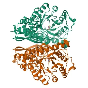
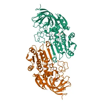
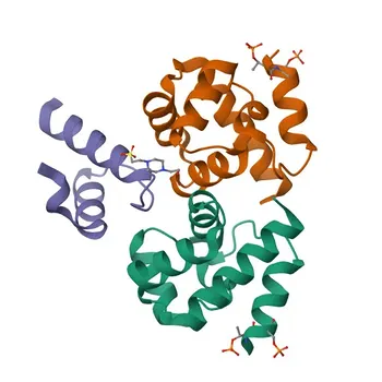
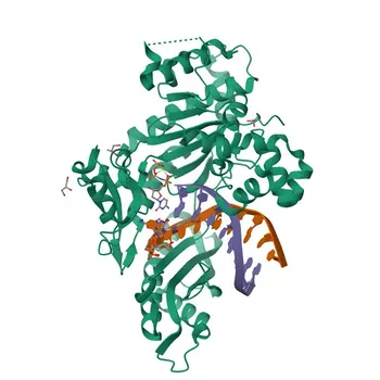
Enzyme-antibody labelling is a cornerstone of modern IVD signal generation. The process involves three pivotal stages: first, selecting and preparing a suitable enzyme and antibody pair; second, chemically conjugating them via activation, bifunctional linkers, or direct coupling; and third, purifying the conjugate from unreacted species, followed by stabilization to maintain long-term activity for consistent colorimetric, fluorimetric, or chemiluminescent readouts.
Robust immunoassay performance hinges on mastering the conjugation chemistry and downstream purification. The goal is not just to link an enzyme to an antibody, but to preserve both enzymatic activity and binding specificity while eliminating any free reactants that cause background noise.
Selecting the Right Enzyme-Antibody System
The entire signal‑generation chain starts with a strategic choice of enzyme and antibody. Not all enzymes are equal, and the intended detection chemistry dictates the selection.
Why Horseradish Peroxidase and Alkaline Phosphatase Dominate
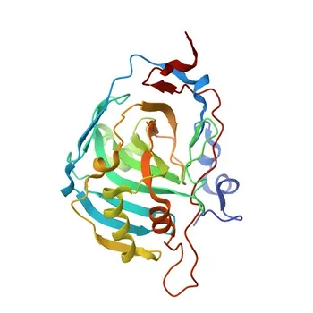
Horseradish peroxidase (HRP) and alkaline phosphatase (AP) are the workhorses because they offer high turnover rates, small size for good conjugate penetration, and compatibility with a wide range of substrates. HRP is favored for its rapid kinetics and cost‑effectiveness in colorimetric and chemiluminescent formats, while AP excels where endogenous peroxidase activity might interfere.
Matching the Enzyme to the Detection Mode
Colorimetric detection often demands enzymes with broad linearity and stable soluble products. Fluorimetric and chemiluminescent readouts require enzymes that amplify the signal without excessive glow. The enzyme’s pH and temperature profile must also align with the assay conditions, ensuring maximal activity when it matters most.
Antibody Quality Is Non‑Negotiable
Even the best enzyme cannot rescue a poor‑quality antibody. The starting antibody must exhibit high specificity, affinity, and minimal background binding. Affinity‑purified polyclonal or recombinant monoclonal antibodies provide the consistency needed for a reliable conjugate, as impurities in the antibody preparation will co‑conjugate and degrade performance.
Conjugation: The Chemistry of Permanent Partnership
The conjugation step is where the enzyme and antibody are chemically linked. The choice of technique directly affects the size, orientation, and activity retention of the final conjugate.
Glycoprotein Activation: Exploiting Enzyme Glycans
Many enzymes, including HRP, are glycoproteins. By oxidizing the sugar moieties with periodate, you generate reactive aldehyde groups. These then form stable Schiff bases with primary amines on the antibody, followed by reduction to secure a covalent bond. This method is gentle on the enzyme’s active site, often preserving high activity.
Bifunctional Linkers: Engineering Precision Bridges
Heterobifunctional crosslinker molecules (e.g., SMCC) allow stepwise, site‑directed conjugation. One reactive group first attaches to the enzyme, while the second is designed to react specifically with amine or sulfhydryl groups on the antibody. This approach minimizes random polymerization and helps maintain the antigen‑binding region’s accessibility, yielding more homogeneous conjugates.
Direct Coupling of Small Molecules
When the enzyme itself is the analyte or a small hapten, direct coupling using carbodiimide chemistry can form a zero‑length bond. However, for antibody labelling, this method is less common because it risks crosslinking the antibody’s binding site. Still, it remains a versatile option for certain low‑molecular‑weight analytes.
Purification: Removing the Noise to Reveal the Signal
Post‑reaction, the mixture contains the desired conjugate, unreacted enzyme, free antibody, and aggregates. Purification is essential—failure here leads to high background and reduced sensitivity.
Gel Filtration: Size‑Based Separation
Gel filtration (size‑exclusion chromatography) quickly separates conjugates based on molecular weight. It is gentle and scalable, efficiently removing excess small crosslinkers and buffer‑exchanging the conjugate. However, it may not fully resolve monomeric conjugate from free enzyme or antibody if their sizes overlap.
Lectin Affinity Chromatography: Exploiting Glycoprotein Specificity
Since HRP is a glycoprotein, lectin columns (like Concanavalin A–Sepharose) can specifically capture the enzyme‑containing fraction. Free antibodies lacking glycans pass through, while the conjugate is eluted with a competing sugar. This method dramatically enriches for enzyme‑labelled antibody, boosting specific activity.
Protein A Affinity Chromatography: Antibody‑Focused Polishing
If the antibody is from a species that binds to Protein A (e.g., rabbit, human IgG), this affinity step can remove free enzyme. The conjugate and free antibody both bind, but subsequent elution can be optimized to collect the active conjugate. Combining Protein A with a prior gel filtration step yields exceptionally pure conjugates.
Stabilization: Preserving Functional Integrity
A highly active conjugate can lose potency within hours without proper stabilization. Long‑term storage demands a formulation that protects both the enzymatic activity and the antibody’s binding capability.
The Role of Stabilizers
Proteins like bovine serum albumin (BSA), along with sugars, polyols, and detergents, crowd the solution and prevent surface denaturation. They also scavenge free radicals and chelate heavy metals that would otherwise poison the enzyme’s active site. Tailored stabilizer cocktails maintain consistent signal over months, even in liquid form.
Activity Retention Over Time
The goal is to ensure the conjugate performs identically from the day it is made to the end of its shelf life. Accelerated stability studies at elevated temperatures can predict real‑time loss, allowing fine‑tuning of buffer pH, salt concentration, and protein load. A well‑stabilized conjugate makes the difference between a reliable IVD kit and one that drifts over time.
Understanding the Trade‑offs
No single workflow is universally optimal. Each step carries inherent compromises that you must weigh against your assay’s requirements.
- Large‑scale vs. research‑grade production: Bulk glycoprotein activation is economical but can generate batch‑to‑batch variability. Bifunctional linkers offer consistency at a higher cost.
- Purity vs. yield: Aggressive purification removes more unreacted species but inevitably sacrifices some active conjugate. Balancing sensitivity against recovery is critical for kit economics.
- Stabilizer interactions: Some stabilizers (e.g., certain polymers) may interfere with the signal readout or increase viscosity. Always validate the final formulation in the complete assay environment.
- Enzyme choice lock‑in: Switching from HRP to AP later in development requires re‑optimizing the entire conjugation‑purification‑stabilization cascade, as the chemistries are not interchangeable.
Making the Right Choice for Your Goal
Your specific immunoassay demands will dictate which path to prioritize. Use these goal‑driven guidelines to focus your efforts.
- If your primary focus is maximizing signal sensitivity: Start with HRP activated via periodate, followed by lectin and Protein A tandem purification, and stabilize with a commercially validated cocktail for chemiluminescent substrates.
- If your primary focus is reducing background noise: Choose a heterobifunctional linker conjugation to ensure consistent antibody orientation, coupled with rigorous gel filtration to exclude aggregates.
- If your primary focus is long‑term stability in a liquid IVD kit: Invest early in stabilizer screening—testing sugars, BSA, and chelators in combination—and monitor enzyme activity decay at multiple temperatures.
- If your primary focus is cost‑effective scale‑up: Favor glycoprotein activation and gel filtration as the core workhorses, accepting a slightly lower purification fold for a more robust and less expensive process.
A meticulously prepared enzyme‑labelled antibody is not just a reagent—it is the engine of signal generation that turns a molecular recognition event into a reliable diagnostic answer.
Summary Table:
| Process Stage | Primary Methods & Chemistries | Main Objective & Key Benefits |
|---|---|---|
| 1. Selection | HRP / AP enzymes; Affinity-purified antibodies | Ensures high kinetic turnover and minimal cross-reactivity |
| 2. Conjugation | Glycoprotein activation (periodate), Bifunctional linkers (SMCC) | Forms covalent bonds while protecting active and binding sites |
| 3. Purification | Gel filtration (SEC), Lectin & Protein A affinity | Eliminates unreacted reagents and aggregates to lower background |
| 4. Stabilization | BSA, polyols, sugars, chelating agents | Protects against denaturation and maintains long-term shelf life |
Accelerate Your IVD Immunoassay Development with CamelBio
Developing high-performance enzyme-antibody conjugates requires precision chemistry and rigorous purification at every step. CamelBio provides diagnostic manufacturers, labs, and research institutes with one-stop access to premium IVD raw materials, custom technical services, and expert consulting—supporting your assay from concept to clinic.
Ready to optimize your assay sensitivity, eliminate background noise, and scale up production? Contact us today to discuss your IVD development needs with our specialists!
Related Products
- Anti-ENO2 Rabbit Polyclonal Antibody for WB, IF/ICC, IP, ELISA - P09104
- Anti-Alcohol Dehydrogenase 1A Monoclonal Antibody for WB, ELISA - P07327
- Anti-Emerin/EMD Polyclonal Antibody for WB, IHC-P, IF/ICC, ELISA - P50402
- Anti-STX1A Polyclonal Antibody for WB, IF/ICC, ELISA - Q16623
- Anti-DNA polymerase eta Polyclonal Antibody for WB - Q9Y253