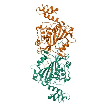
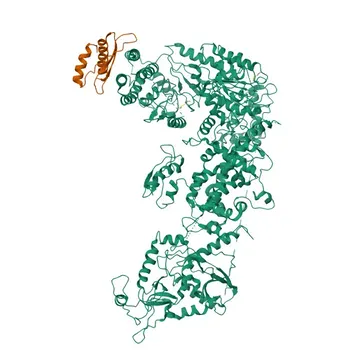
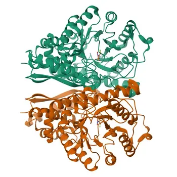
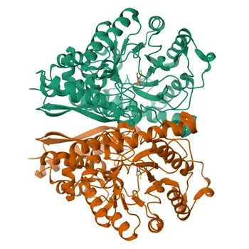
The mechanism is deceptively simple. Restriction endonucleases—primarily Type II enzymes—recognize specific palindromic DNA sequences (such as 5′-GAATTC-3′ for EcoRI) and cleave both strands at precisely defined phosphodiester bonds. In an RFLP diagnostic assay, a single nucleotide mutation that alters this recognition site changes the number and size of the resulting DNA fragments, creating a unique pattern on a gel. The true complexity, and the diagnostic power, lie in selecting an enzyme that performs this surgery with absolute fidelity every time.
The reliability of an RFLP-based diagnostic hinges on one factor: the enzyme’s unwavering specificity. By choosing restriction endonucleases with high site fidelity, consistent batch‑to‑batch activity, and negligible star activity, you transform a simple digestion into a reproducible, high‑confidence genetic test.
The Molecular Logic of Restriction Endonucleases
A Precision Lock‑and‑Key Recognition
Type II restriction enzymes bind to double‑stranded DNA and scan for a short, often symmetrical recognition sequence. Once bound, the enzyme induces a conformational change that positions catalytic residues to simultaneously break the phosphodiester backbone of each strand. This lock‑and‑key mechanism is what makes the enzyme so predictable—and so valuable for diagnostics.
How a Cut Site Becomes a Diagnostic Signal
In RFLP analysis, the enzyme digests the sample DNA wherever its recognition sequence appears. If a mutation or polymorphism abolishes a cut site or creates a new one, the fragment pattern changes. After separation by gel electrophoresis, these fragment length differences become visible as altered banding patterns. This principle allows laboratories to detect genetic variants, identify pathogen subtypes, and perform forensic matches without sequencing.
Sticky Ends, Blunt Ends, and Fragment Readout
Cleavage can produce sticky (cohesive) ends with short overhangs or blunt ends with no overhang. While end type is critical for downstream cloning, in RFLP the only parameter that matters is the length of the fragment generated. Enzymes that yield clean, predictable cuts—regardless of end morphology—are what enable precise sizing and accurate genotype calling.
Selecting the Right Enzyme: Critical Factors for Diagnostic Reliability
Minimizing Star Activity for Unambiguous Results
Star activity is the enzyme’s tendency to cleave at degenerate, non‑canonical sites under sub‑optimal conditions (e.g., high glycerol, incorrect buffer, excess enzyme). Even a single off‑target cut introduces false fragments that can mimic a polymorphism. Diagnostic‑grade enzymes are rigorously optimized and tested to suppress star activity, ensuring that every band on the gel corresponds to the intended recognition site.
Guaranteeing Consistent Cleavage Efficiency
Lot‑to‑lot variation in enzyme activity is a silent assay killer. Slight drops in efficiency lead to partial digestions, producing faint or missing bands that are easily mistaken for genetic absence. For clinical diagnostics, manufacturers must source enzymes that come with validated activity certificates and demonstrate identical kinetics across independent production batches.
Purity as a Shield Against Non‑Specific Nucleases
Commercial enzyme preparations can harbor contaminating exonucleases or phosphatases that nibble at fragment ends or degrade DNA. Such degradation distorts fragment sizes and reduces overall signal intensity. High‑purity restriction endonucleases, often processed through multiple orthogonal chromatography steps, eliminate these background activities and guarantee that every fragment measured is a direct result of site‑specific cleavage.
Matching Cut Frequency to the Diagnostic Target
The frequency of the recognition sequence in your target genome must drive enzyme choice. For high‑resolution genotyping of complex eukaryotic genomes, enzymes that recognize short, 4‑base pair sequences (e.g., HaeIII at GGCC or HinfI at GANTC) cut frequently, yielding many small fragments that maximize the chance of revealing a polymorphism. For targeted plasmid or pathogen verification, a rarer cutter with a longer recognition site might be more appropriate to avoid unmanageable fragment complexity.
Understanding the Trade‑offs
Even the best restriction enzymes come with inherent compromises. High‑frequency cutters increase resolution but also generate a dense, overlapping band pattern that can be difficult to interpret without sophisticated imaging and precise size markers. This complexity demands standardized reference ladders and control DNA to calibrate each gel run.
Purity and lot‑consistency elevate cost. Diagnostic‑grade enzymes are more expensive than research‑grade alternatives because of the additional quality control. For IVD manufacturers, however, this cost must be weighed against the risk of an equivocal clinical result. Cutting corners on enzyme quality can lead to false inclusion or exclusion calls that undermine diagnostic credibility.
Finally, suppressing star activity often requires strict adherence to the manufacturer’s exact buffer system and incubation time. Deviations intended to speed up workflows or reduce reagent costs can inadvertently relax specificity, reintroducing the very ambiguity you sought to eliminate. Robust assay design therefore means building the protocol around the enzyme’s optimal condition, not the other way around.
How to Build a Robust RFLP Assay
Your choice of restriction endonuclease must align with your diagnostic goal. Use these criteria to guide selection:
- If your primary focus is high‑resolution genotyping (e.g., forensic matching or microbial strain typing): Select a high‑frequency cutter such as HinfI or HaeIII that produces many small fragments, increasing the probability that a polymorphism falls exactly at a recognition site.
- If your primary focus is clinical repeatability and regulatory compliance: Prioritize diagnostic‑grade enzymes with documented lot‑to‑lot consistency, negligible star activity, and purity that eliminates non‑specific nucleases. Every band must be real, every run must be reproducible.
- If your primary focus is cost‑sensitive assay development: Start with well‑characterized, widely available enzymes like EcoRI, but rigorously validate buffer conditions and enzyme stability under your exact sample preparation workflow. Never assume that research‑grade purity will perform identically in a clinical matrix.
Selecting the right restriction endonuclease isn’t just about cutting DNA—it’s about building a foundation of trust in every diagnostic result.
Summary Table:
| Selection Criterion | Diagnostic Impact | Key Requirement / Solution |
|---|---|---|
| Star Activity Suppression | Prevents off-target cuts that cause false bands | Optimized buffer systems & diagnostic-grade fidelity |
| Lot-to-Lot Consistency | Prevents partial digestions and assay failure | Validated activity certificates & stringent QC |
| High Enzyme Purity | Protects target DNA from non-specific degradation | Multi-step chromatography with zero exonuclease contamination |
| Recognition Sequence Length | Balances fragment resolution and band pattern complexity | Short 4-bp cutters for genotyping; longer cutters for targeted assays |
Optimize Your Molecular Diagnostic Assays with High-Fidelity Enzymes
Reliable RFLP and genetic assays depend on uncompromising enzyme purity, lot-to-lot consistency, and zero star activity. CamelBio provides diagnostic manufacturers, labs, and research institutes with one-stop access to IVD raw materials, technical services, and consulting—covering every stage from concept to clinic.
Whether you are scaling IVD production or developing custom molecular diagnostic protocols, our team is ready to support your success. Contact CamelBio today to request technical specifications and samples!
Related Products
- Anti-Endo G Rabbit Monoclonal Antibody for WB, ELISA - Q14249
- Anti-ENDOGL1/ENGL Rabbit Monoclonal Antibody for WB and ELISA - Q9Y2C4
- Anti-DICER1 Polyclonal Antibody for IF/ICC, ELISA - Q9UPY3
- Anti-CD39/ENTPD1 Monoclonal Antibody for FC, ELISA - P49961
- Anti-RNASE2 Rabbit Polyclonal Antibody for WB - O35291