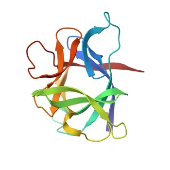
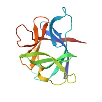
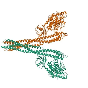
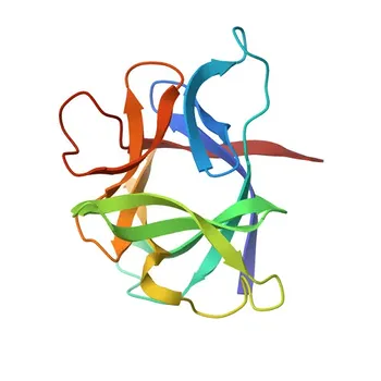
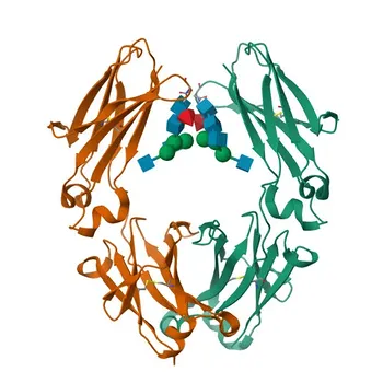
False positives in anti-cardiolipin and anti-β2 glycoprotein I assays are far more than a technical nuisance—they can trigger a lifelong diagnosis of antiphospholipid syndrome and its associated thrombotic risks. The most powerful mitigation is to design the kit around the true autoimmune target: the β2 glycoprotein I (β2 GPI) / phospholipid complex. By using purified β2 GPI as a co-antigen alongside cardiolipin, you eliminate reactivity from transient, infection‑driven antibodies that bind cardiolipin alone. You must also enable separate, quantitative detection of IgG and IgM isotypes and embed a mandatory 12‑week confirmatory retesting protocol to allow transient titers to wane, ensuring only chronic autoimmune antibodies are flagged.
The path to reliable phospholipid antibody diagnosis lies in mimicking the disease’s molecular reality. That means shifting from cardiolipin‑only antigens to the β2 GPI‑dependent complex, demanding antibody class discrimination, and fortifying the entire assay with stringent blocking, wash optimization, and raw‑material validation to silence non‑specific noise. The ultimate safeguard is a re‑test window that time‑filters infection‑derived false positives from true autoimmune disease.
Understanding the Source of False Positives in Phospholipid Antibody Testing
The classic cardiolipin ELISA can betray the clinician because it cannot distinguish autoimmune antibodies from benign, infection‑induced ones.
Transient Antibodies During Acute Infection
Many common infections—especially syphilis (which famously generates cardiolipin reactivity in VDRL tests)—provoke transient anti‑cardiolipin antibodies.
These antibodies often target the phospholipid directly, without requiring a protein co‑factor.
They are typically of the IgM isotype and fade within a few weeks to months after the infection clears.
The True Autoimmune Target
In antiphospholipid syndrome, pathogenic antibodies do not bind cardiolipin in isolation.
They recognize conformational epitopes on β2 GPI that become exposed when the protein binds to negatively charged phospholipid surfaces.
Failing to present this complex in the assay invites non‑specific signals from infection‑related antibodies, undermining diagnostic specificity.
Designing the Antigenic Target: Putting β2 GPI at the Center
The single most important design decision is to build the assay around the β2 GPI‑dependent cardiolipin complex.
Purified β2 GPI as a Co‑Antigen
Use highly purified human or recombinant β2 GPI as an essential co‑reactant alongside an anionic phospholipid preparation.
When the cardiolipin surface is pre‑incubated with β2 GPI, only antibodies that require this co‑factor will bind.
This immediately filters out direct phospholipid‑reactive antibodies that are common after infections.
Recombinant and Synthetic Antigens
Opt for recombinant β2 GPI domain‑specific constructs or synthetic peptides if you need to refine epitope specificity even further.
Such designed antigens eliminate batch‑to‑batch variability and can reduce cross‑reactivity with other serum proteins that might mimic the natural co‑factor.
Antibody Class‑Specific Detection: Cutting Through the Noise
Detecting total immunoglobulins lumps together transient IgM with persistent IgG, inflating false‑positive rates.
Separate IgG and IgM Channels
Design the kit to individually measure IgG and IgM anti‑β2 GPI‑cardiolipin antibodies.
Transient infectious antibodies are heavily biased toward IgM; a purely IgG‑positive result is far more suggestive of a chronic autoimmune process.
Quantifying each isotype also enables clinicians to apply the latest classification criteria that demand medium‑to‑high titers.
Eliminating Fc‑Mediated Interactions
Use antibody fragments—Fab or F(ab')₂—for both capture and detection reagents.
Removing the Fc region abolishes binding sites for heterophilic antibodies and human anti‑mouse antibodies (HAMA) that could bridge capture and detection antibodies in the absence of the target analyte.
This step alone dramatically reduces one of the most common matrix‑interference false‑positive mechanisms.
Engineering Out Non‑Specific Binding and Interfering Substances
Even with a perfect antigen, physical and chemical noise can generate a false signal.
Robust Blocking and Buffer Chemistry
Formulate the assay diluent and wash buffer to suppress ionic and hydrophobic interactions.
High ionic strength buffers disrupt weak electrostatic binding, while a blend of non‑ionic detergents and non‑relevant blocking proteins (e.g., bovine serum albumin, casein) saturates sticky surfaces.
If background persists, escalate the wash buffer pH up to 12 to shear off loosely adherent immunoglobulins without stripping specifically bound antibodies.
Neutralizing Heterophile and Anti‑Animal Antibodies
Include active heterophile blocking reagents, non‑immune mouse IgG, or nonimmune animal sera directly in the sample diluent.
These additives scavenge endogenous HAMA and heterophile antibodies before they can cross‑link assay components.
For sandwich formats, this is non‑negotiable; without it, even healthy samples can produce a signal.
Raw Material Screening and Validation
Screen every antibody lot and antigen preparation against extensive panels of potential cross‑reactants: other phospholipid‑binding proteins, structurally related co‑factor‑dependent antibodies, and common infectious sera.
Confirm that the detection antibody pair delivers a sharp dose‑response curve with minimal off‑target binding near the clinical cut‑off.
Only materials that demonstrate restricted reactivity and linear dilution recovery should be released for kit formulation.
Incorporating the 12‑Week Confirmatory Protocol
No matter how well the assay is engineered, the clinical context remains the ultimate arbiter.
Mandatory Retesting After Three Months
The kit’s instructions for use must explicitly require a repeat test 12 weeks after an initial positive result.
This interval allows transient, infection‑driven titers to decay, transforming a potentially false‑positive snapshot into a sustained, persistent antibody profile.
Autoimmune antiphospholipid antibodies, in contrast, remain stable or rise during this period.
Supporting Dilution‑Linearity Verification
During validation, include serial dilution studies in the kit’s performance documentation.
If a positive sample shows non‑linear recovery upon dilution, it strongly suggests matrix interference rather than a true antibody response.
Laboratories can then flag such samples for re‑testing on an alternative platform or for confirmatory functional assays.
Understanding the Trade‑offs
No single mitigation strategy comes without compromise. A transparent view of these tensions builds trust with end‑users.
Specificity vs. Sensitivity
Requiring β2 GPI dependency and high‑titer IgG positivity maximizes specificity but may miss rare patients whose pathogenic antibodies recognize cardiolipin through a different co‑factor (e.g., prothrombin).
If the goal is a screening kit, you may need to accept a slightly lower specificity to capture all potential cases, then refer clearly for confirmatory testing.
The 12‑Week Window as a Diagnostic Delay
Mandating a three‑month confirmatory gap introduces a deliberate wait.
For a patient with an acute thrombotic event, this can feel like an unacceptable delay.
Clear communication in the kit insert and physician‑facing materials must explain that immediate treatment decisions should never rely on a single positive anti‑phospholipid test.
Complexity and Cost
Adding purified β2 GPI, separate IgG/IgM channels, and extensive blockers increases manufacturing complexity and cost per test.
You must weigh this against the clinical and reputational risk of false‑positive results.
In specialized autoimmune testing, laboratories typically accept a higher cost in exchange for diagnostic confidence.
Making the Right Choice for Your Kit
Align your design choices with the exact clinical problem you intend to solve.
- If your primary focus is maximum diagnostic specificity for antiphospholipid syndrome: Build a β2 GPI‑dependent cardiolipin assay with distinct IgG and IgM quantification, Fab‑based reagents, and a hard‑coded 12‑week retesting requirement.
- If your primary focus is a screening tool that must not miss any potential case: Consider including a cardiolipin‑only test channel in parallel with the β2 GPI‑dependent channel, but flag all isolated cardiolipin positives as requiring confirmation and correlate clearly with infection history.
- If your primary focus is ease of use in resource‑limited settings: Prioritize robust blocking chemistry and heterophile neutralization in a single‑well format with a single IgG cut‑off, and include an insert that clearly trains users to repeat any positive with a fresh sample after 12 weeks.
A false‑positive result in this field is not just a number; it is a patient living under a disease label. By engineering your kit to recapitulate the true autoimmune interaction and then reinforcing that design with disciplined clinical validation, you turn a notorious diagnostic pitfall into a foundation of trust.
Summary Table:
| Mitigation Strategy | Mechanism of Action | Primary Clinical & Technical Benefit |
|---|---|---|
| β2 GPI Co-Antigen Presentation | Presents β2 GPI/cardiolipin complex rather than isolated cardiolipin | Eliminates non-specific, infection-driven transient antibody binding |
| Separate IgG & IgM Channels | Quantitative discrimination of antibody isotypes | Distinguishes chronic autoimmune antibodies (IgG) from transient IgM |
| Fab / F(ab')₂ Reagents | Cleaves Fc region from capture and detection antibodies | Prevents HAMA and heterophile antibody cross-linking interferences |
| Optimized Buffer Chemistry | High ionic strength, non-ionic detergents, and high pH washes | Disrupts weak non-specific electrostatic and hydrophobic interactions |
| 12-Week Retest Requirement | Embedded confirmatory window in kit instructions | Filters out decaying post-infectious antibodies from persistent autoimmune cases |
Elevate Your Immunoassay Specificity with CamelBio
Eliminating false positives requires high-quality antigens, optimized reagents, and expert assay engineering. At CamelBio, we provide diagnostic manufacturers, clinical labs, and research institutes with one-stop access to premium IVD raw materials, technical services, and consulting—covering every stage from concept to clinic.
Whether you need high-purity β2 GPI antigens, Fab fragment reagents, or custom buffer formulations to eliminate matrix interference, our experts are here to help.
Contact CamelBio Today to optimize your assay performance and accelerate your path to market!
Related Products
- Anti-CMIP Polyclonal Antibody for WB, IHC-P, ELISA - Q8IY22
- Anti-LCAT Monoclonal Antibody for WB, ELISA - P04180
- Anti-GIPC1 Rabbit Polyclonal Antibody for WB, ELISA - O14908
- Anti-hCG beta Monoclonal Antibody for WB, IF-P, IHC-P, ELISA - P0DN86
- IL2 Rabbit Monoclonal Antibody for WB, ELISA - P60568
People Also Ask
- What sample matrix challenges must nucleic acid extraction raw materials resolve to prevent PCR inhibition?
- How do nanomaterial multilabeling strategies overcome 1:1 ELISA limits? Boost IVD Sensitivity Up to 1000x
- Why prioritize kinetic binding over affinity in lateral flow test strips? Optimize LFA antibody selection
- How do nitrocellulose coatings & colloidal carbon enable multiplex IVD assays? Key Benefits
- How to optimize chemiluminescent assay reagents for ROS direct detection? Step-by-Step Guide