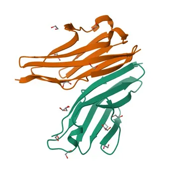
The short answer is that morphine-targeted antibodies simply don’t recognize oxycodone well enough. Standard opiate screening immunoassays are built on antibodies raised against morphine. Because oxycodone has a structurally distinct backbone, these reagents often exhibit poor cross-reactivity, causing dangerously high rates of false-negative results. Diagnostic manufacturers must therefore replace broad-spectrum opiate raw materials with dedicated oxycodone-specific antibodies and matched protein conjugates to achieve the sensitivity and accuracy modern pain management testing demands.
The core problem is a molecular mismatch. Morphine-derived antibodies were designed for a different target, leaving semisynthetic opioids like oxycodone analytically invisible. The only reliable fix is to engineer separate immunoassay channels around antibodies that see oxycodone’s unique epitope – no amount of panel tweaking can compensate for a fundamentally wrong recognition surface.
The Structural Blind Spot: Why Morphine Antibodies Miss Oxycodone
Historical Roots in Heroin Detection
Opiate screening immunoassays were originally developed to catch heroin use. The antibodies were raised against morphine, heroin’s primary metabolite, because that was the most relevant public health target.
This legacy means the binding pockets of these antibodies are shaped to fit morphine’s 5‑membered nitrogen ring and phenolic hydroxyl group. When faced with a semisynthetic derivative like oxycodone, the fit is often so poor that even high analyte concentrations fail to trigger a positive signal.
Chemical Differences Between Morphine and Oxycodone
Morphine and oxycodone are both opiates, but their structural differences are decisive. Oxycodone replaces the phenolic –OH with a methoxy group, adds a ketone at C‑6, and saturates the C‑7–C‑8 double bond.
These modifications subtly, yet profoundly, alter the molecule’s electron cloud and three‑dimensional shape. An antibody raised against morphine sees a foreign surface where key hydrogen‑bond donors and hydrophobic contacts no longer align.
Cross‑Reactivity Profiles: Numbers That Matter
For a screening assay to be clinically useful, an antibody must generate a strong signal at or below the target’s cutoff concentration. With oxycodone, morphine‑directed antibodies frequently deliver cross‑reactivity below 5% of the homologous morphine response.
This means a patient taking a high therapeutic dose of oxycodone can repeatedly test negative. The assay simply doesn’t recognize the drug that should be driving the signal, turning a compliance screen into a false‑negative liability.
The Clinical Risk of False‑Negative Opiate Screens
Undermining Pain Management Compliance
In pain management programs, clinicians rely on urine drug testing to confirm that a patient is actually taking prescribed oxycodone and not diverting it. A false‑negative result wrongly suggests non‑compliance, potentially leading to incorrect accusations, loss of trust, or inappropriate discharge from a care program.
When the screening antibody cannot see the prescribed drug, the entire therapeutic contract becomes based on incomplete data.
The Cascade Effect on Confirmatory Testing
Many laboratories use immunoassay screens as a gatekeeper: only screen‑positive samples proceed to more expensive LC‑MS/MS confirmation. If the screen is blind to oxycodone, the confirmatory step is never triggered.
The patient’s oxycodone goes undetected, even though the drug is present at therapeutic levels. The result is a laboratory report that says “negative” for a drug the patient is definitely taking — a systemic failure of the testing protocol.
Engineering the Solution: A Dedicated Oxycodone Immunoassay
Antibody Specificity: Starting with a Clean Slate
The only way to close the detection gap is to develop an immunoassay around an antibody raised specifically against oxycodone. This antibody must be immunized with a hapten that preserves oxycodone’s full ring system and functional groups, presenting the exact molecular surface that needs to be recognized.
A well‑designed oxycodone‑specific antibody will show greater than 90% cross‑reactivity with the parent drug and minimal interference from morphine, codeine, or their glucuronide metabolites. This specificity is not a luxury; it is the analytical foundation of a trustworthy result.
The Role of Matched Protein Conjugates
Antibody specificity alone is not enough. The protein conjugate used in the assay — the competing binder — must be carefully matched to the antibody’s binding cleft.
If the conjugate’s linker arm masks the same structural features that differentiate oxycodone from morphine, the assay loses its discriminatory power. A matched conjugate strategy ensures that the competitive displacement exactly reflects the target analyte’s presence, preserving both sensitivity and selectivity.
Assay Design Considerations for Sensitivity and Cutoff
A dedicated oxycodone assay must be tuned to the clinically relevant cutoff, typically 100–300 ng/mL. Developers need to select an antibody‑conjugate pair that delivers a steep dose‑response curve right at that threshold.
Factors like pH, ionic strength, and incubation time can shift cross‑reactivity patterns. Rigorous optimisation against oxycodone metabolites (e.g., noroxycodone, oxymorphone) is also essential to avoid over‑ or under‑estimating the parent drug and to align with medical guidelines.
Understanding the Trade‑offs
Increased Reagent Complexity and Cost
Adding an oxycodone‑specific channel means more raw materials, more QC controls, and a higher manufacturing burden. A multiplex panel that separates oxycodone from classical opiates will always be more complex than a one‑size‑fits‑all morphine screen.
Manufacturers must weigh the added cost against the risk of undetected non‑compliance — a calculation that, in regulated clinical markets, almost always favours the dedicated approach.
Potential for Antibody Lot Variability
Every monoclonal antibody lot can exhibit slight affinity shifts. For a dedicated oxycodone assay, even a 2–3% drift in cross‑reactivity can move the effective cutoff and affect clinical classification.
Robust lot‑to‑lot validation and monitoring of binding kinetics are essential. Without these controls, the assay can drift from “specific for oxycodone” to “partially blind to oxycodone” over successive production runs.
Balancing Panel Breadth vs. Single‑Target Accuracy
A panel capable of detecting dozens of opioids with a single sample is attractive. However, squeezing oxycodone detection into a broad‑spectrum opiate channel inevitably sacrifices accuracy.
The engineering truth is that high‑specificity detection of semisynthetic opioids demands its own reagent channel. Attempting to stretch a morphine‑derived antibody to cover oxycodone will degrade the performance of both detections.
Making the Right Choice for Your Diagnostic Platform
Below are the practical pathways for manufacturers, based on the clinical problem they are solving.
- If your primary focus is eliminating false negatives in pain management compliance: Invest in a dedicated oxycodone immunoassay with a highly specific antibody, and validate cross‑reactivity against common metabolites and co‑prescribed drugs.
- If your primary focus is developing a multi‑analyte opiate panel: Design separate reagent channels for oxycodone and classical opiates rather than forcing one morphine‑based antibody to cover all targets, even if per‑test costs increase.
- If your primary focus is meeting regulatory guidelines for drug‑of‑abuse screening: Provide clear performance data showing negligible interference from morphine‑type opiates and robust detection of oxycodone at clinically mandated cutoffs, with traceable lot‑to‑lot consistency studies.
Choosing the right antibody and conjugate from the beginning transforms a screening shortfall into a reliable, high‑trust diagnostic asset.
Summary Table:
| Parameter / Aspect | Morphine-Targeted Reagents | Dedicated Oxycodone Reagents |
|---|---|---|
| Epitope Target | Morphine / Heroin primary metabolites | Unique oxycodone methoxy & C-6 ketone structure |
| Oxycodone Cross-Reactivity | Poor (< 5%) | High (> 90%) |
| Clinical Impact | High risk of false-negative screens | Accurate pain management compliance testing |
| Assay Engineering | Broad-spectrum compromise | Dedicated reagent channel + matched protein conjugate |
Upgrade Your Oxycodone Immunoassay Performance with CamelBio
Eliminate false-negative risks and optimize your drug-of-abuse testing panels with high-specificity antibodies and custom-matched protein conjugates. CamelBio provides diagnostic manufacturers, labs, and research institutes with one-stop access to high-quality IVD raw materials, technical services, and consulting—covering every stage from concept to clinic.
Ready to enhance your diagnostic assay's accuracy and reliability? Contact CamelBio today to consult with our IVD raw material experts!
Related Products
- Anti-CD4 Rabbit PolymAb® for WB, IF-P, IHC-P, ELISA - P06332
- Anti-LCAT Monoclonal Antibody for WB, ELISA - P04180
- Anti-GIPC1 Rabbit Polyclonal Antibody for WB, ELISA - O14908
- Anti-FOXO1/FOXO3A/FOXO4 Polyclonal Antibody for WB, IHC-P, ELISA - Q12778 / O43524 / P98177
- Anti-HOOC4 Polyclonal Antibody for WB, ELISA - P09017