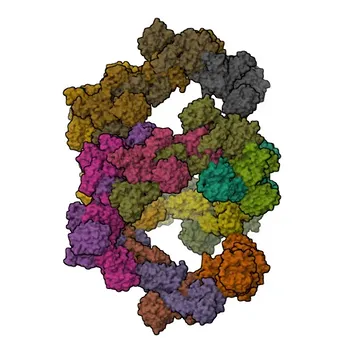
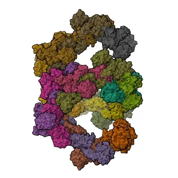
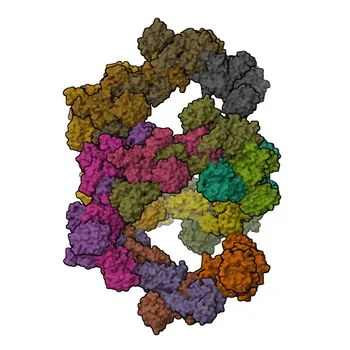
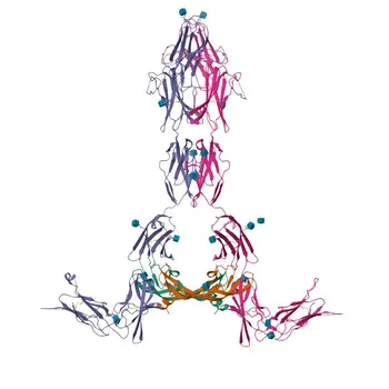
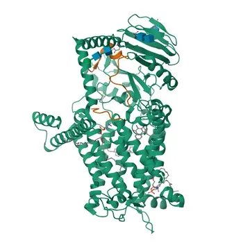
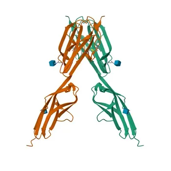
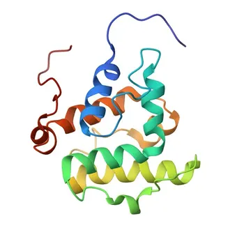
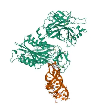
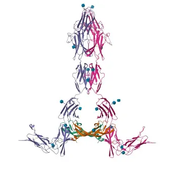
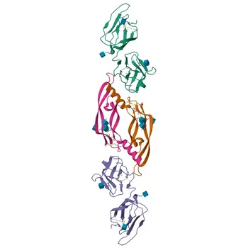
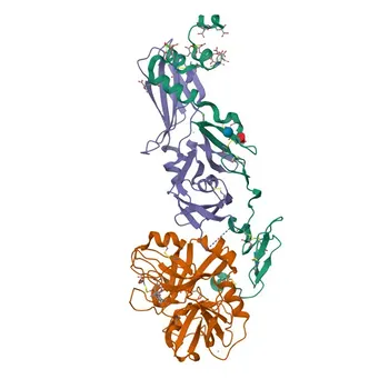
Selecting raw materials for VWF immunoassay kits is not about sourcing any VWF protein—it’s about preserving the precise structural domains and multimeric integrity that define all of its hemostatic functions.
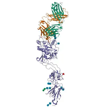
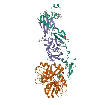
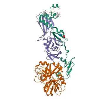
Diagnostic manufacturers must evaluate whether candidate antibodies, recombinant proteins, and calibrator plasma retain the native conformation of the A1 domain (platelet GPIb binding), the A3 domain (collagen binding), and the D’/D3 domains (Factor VIII binding). Simultaneously, they must confirm that the material’s multimer size distribution mirrors physiological VWF, including high-molecular-weight multimers (HMWM) that are critical for accurate functional activity measurements and for distinguishing von Willebrand disease subtypes.
The core challenge: Functional VWF assays do not just measure mass—they measure biological activity that depends on large, flexible multimers and correctly folded sub-domains. Raw materials must therefore be qualified not only for antigenic content but for domain-specific reactivity, multimer pattern integrity, and the preservation of shear-dependent or collagen-binding function. A failure on any of these fronts can conceal type 2 VWD variants or produce misleading Factor VIII binding results.
The Multidomain Architecture: Why Structure Dictates Function
VWF’s functional portfolio is structured into distinct domains, each responsible for a different binding interaction. An immunoassay’s ability to capture these activities depends entirely on the raw material’s epitope presentation.
The A1 Domain and Platelet Adhesion
The A1 domain is the docking site for the platelet receptor GPIb. This interaction is the very basis of primary hemostasis under high shear—think of it as the initial tether that catches a platelet flowing past a wound.
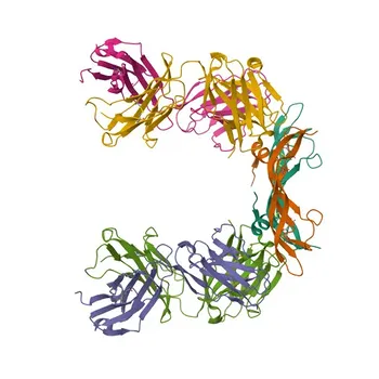
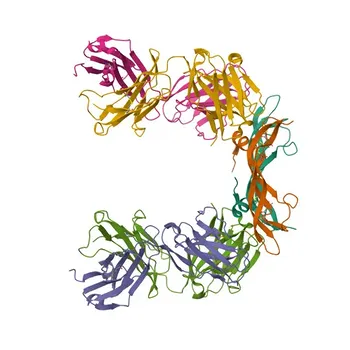
For diagnostic kits measuring VWF platelet-binding function (e.g., VWF:RCo or its modern replacements), the antibody or receptor used in the reagent must target the GPIb-binding epitope within the A1 domain in its native, undamaged conformation. Even slight denaturation can abolish this binding, making a patient sample appear deficient in functional VWF when it is not. Monoclonal antibodies selected for latex agglutination assays (VWF:Ab) are specifically directed at this functional patch, bypassing the need for ristocetin but demanding that the epitope be intact on the calibrator and sample alike.
The A3 Domain and Collagen Anchoring
The A3 domain binds to subendothelial collagen (types I and III). This anchors VWF to the vessel wall, converting it from a soluble plasma protein into a surface on which platelets can accumulate.
In collagen-binding assays (VWF:CB) , the solid-phase capture material is not an antibody but collagen itself. Here, the raw material’s structural property—the triple-helical integrity of collagen—becomes the critical parameter. Collagen purified from equine, bovine, or human tendon must retain its native helical structure; otherwise, it will not engage the A3 domain properly. This is especially important because VWF:CB is notably sensitive to high-molecular-weight multimers, which have the strongest collagen binding. Loss of collagen helicity leads to a drop in signal for HMWM, potentially missing Type 2A or 2B VWD.
The D’/D3 Domains and Factor VIII Protection
The D’/D3 domains at the N-terminus form a non-covalent binding pocket for Factor VIII. This is a distinct structural job: VWF acts as a protective chaperone that extends FVIII’s half-life in circulation from minutes to hours.
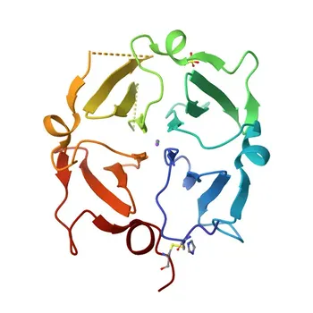
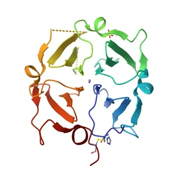
Immunoassay kits that assess VWF-Factor VIII binding capacity or that rely on recombinant VWF fragments as capture agents must preserve the correct folding of these D’/D3 domains. An unfolded or truncated variant might still be recognized by an anti-VWF antibody in a total antigen assay but will fail to bind FVIII, giving a falsely low functional readout. Raw material vendors must demonstrate that their recombinant VWF retains intact FVIII-binding conformation, not just antigenic mass.
The Multimer Pattern: Size is a Functional Unit
VWF exists as a series of multimers from dimers up to ultra-large forms. The hemostatic potency is not uniform—the largest multimers are the most efficient at both platelet capture and collagen binding.
ADAMTS13 Cleavage and the HMWM Imperative
In the body, the protease ADAMTS13 cleaves newly secreted ultra-large VWF multimers into the circulating spectrum of sizes. This cleavage is specific and physiological. When sourcing purified VWF substrate proteins for activity kits, manufacturers need to verify that the multimer distribution includes a representative proportion of HMWM.
If the raw material has been excessively degraded or consists mainly of small multimers, it will underperform in VWF:RCo, VWF:GPIbR, or VWF:CB assays. Even more critically, a calibrator with a poor HMWM profile cannot differentiate Type 2A or 2B VWD (where HMWM are selectively missing) from a true quantitative deficiency. The multimer pattern of the calibrator directly impacts diagnostic specificity.
Assay Format Dictates Which Properties to Prioritize
Different immunoassay strategies put different stressors on the raw material. The choice of format determines which structural features must be explicitly validated.
From Classic Ristocetin to Recombinant Receptor Formats
Traditional VWF:RCo assays depend on whole or lyophilized platelets, which are inherently variable. Modern immuno-turbidimetric alternatives replace them with defined raw materials:
- Monoclonal antibody-based formats (VWF:Ab): Coat latex microparticles with a monoclonal antibody specific to the A1 domain’s GPIb-binding site. In this reagent, the sole structural requirement is that the antibody’s paratope correctly recognizes the native GPIb-binding epitope. Any distortion of this epitope on calibrators or controls will cause an under-recovery.
- Recombinant wild-type GPIb assays (VWF:GPIbR): Immobilize wild-type GPIb receptor fragments and use ristocetin as an agonist. The recombinant GPIb must fold correctly to bind VWF only in the presence of ristocetin. Calibrator VWF must still contain large multimers, because ristocetin-induced binding is highly multimer-size dependent.
- Gain-of-function mutant GPIb assays (VWF:GPIbM): Use engineered GPIb mutants that bind VWF without ristocetin. These assays remove the agonist variable but still require intact A1 domain conformation and a full multimer profile on the VWF substrate.
Collagen-Binding Assays: Material-Specific Demands
For VWF:CB ELISAs, the critical raw material is the coated collagen itself. Developers must evaluate:
- Collagen type selection: Types I and III contain the principal VWF binding sites.
- Source and purity: The collagen must be purified without denaturing its triple-helical conformation.
- Preferential HMWM sensitivity: Assays using this collagen capture are naturally weighted toward HMWM. This is advantageous for detecting qualitative defects but also means that a calibrator lacking HMWM will compress the assay’s dynamic range.
Antibody Selection: Epitope Integrity is the Limiting Factor
Immunoassay specificity and sensitivity ultimately rest on the intrinsic properties of the antibody raw material. For VWF, the choices fall into distinct categories with direct functional consequences.
Polyclonal antibodies can provide broad reactivity across multiple VWF domains and multimers, but they are inherently variable lot-to-lot and may contain irrelevant reactivities. For most high-performance coagulation assays, monoclonal antibodies are preferred because they can be targeted precisely to the functional domain of interest—A1, A3, or D’/D3. The key evaluation is that the monoclonal’s epitope is conformation-dependent and preserved in the final reagent formulation.
Recombinant antibody fragments offer the highest lot-to-lot consistency but still require functional validation: does the recombinant antibody recognize the native VWF domain under the assay’s buffer conditions? A failure here leads to increased non‑specific binding and poor low‑end sensitivity, especially below 30 U/dL where accurate VWD classification lives.
Reagent Formulation: Preserving Structure Long-Term
Selecting an excellent antibody or collagen is only half the battle. The formulation environment—diluents, coupling chemistry, and solid‑phase overcoats—can preserve or destroy the functional structures you’ve chosen.
Coupling of antibodies to latex microparticles can sterically obscure the paratope if sites are randomly crosslinked. Directed coupling through carbohydrate moieties or recombinant handles can leave the antigen‑binding site fully accessible. Diluents containing stabilizing proteins, optimized salts, and low levels of detergent are essential to maintain native VWF multimer structure in calibrator plasmas and to prevent HMWM from precipitating out of solution. Even the solid‑phase overcoat (e.g., BSA, casein) must be rigorously optimized; an insufficient block leaves exposed hydrophobic surfaces that can trap HMWM aggregates and create false‑positive signals.
Understanding the Trade-offs
No single raw material strategy is perfect. There are inherent trade-offs that diagnostic developers must navigate.
Sensitivity vs. specificity in collagen‑binding assays: Collagen surfaces that are hyper‑sensitive to HMWM may also show excessive signal drift with mildly degraded samples, leading to borderline classification of Type 1 vs. Type 2 VWD. You must balance the collagen’s binding avidity so that it discriminates without over‑calling.
Monoclonal precision vs. multimer‑biased detection: Monoclonal antibodies against a single A1 epitope give exceptional reproducibility but can underestimate functional defects when the defect lies outside that precise epitope (e.g., in the A3 domain). A panel that relies on multiple domain‑specific monoclonals or complementary functional assays is often necessary.
Ristocetin‑dependent vs. ristocetin‑independent assays: Ristocetin‑based systems require the raw material to function in the presence of an antibiotic agonist, which can precipitate plasma proteins and artificially influence the signal. The gain‑of‑function mutant GPIb approach removes this variable but demands that the engineered receptor maintains specificity; any loss of discrimination can cause spontaneous binding to VWF variants that should not be active.
Making the Right Choice for Your Diagnostic Goal
The specific assay you are building determines which structural and functional properties must be non-negotiable in your raw materials.
- If your primary focus is a screening assay for VWD type‑1 and type‑3: Prioritize raw materials with proven broad multimer reactivity and a polyclonal or domain‑spanning monoclonal cocktail. Conformation‑preserved pooled plasma calibrators with certified HMWM content are essential to avoid falsely low antigen values.
- If your primary focus is discriminating type‑2A or type‑2B VWD: Demand collagen with native triple‑helical structure and calibrators that clearly demonstrate a HMWM‑selective signal. Validate that your antibody or GPIb reagent fails to bind ultra‑large forms in the absence of agonist if using a ristocetin‑free format.
- If your primary focus is a VWF‑Factor VIII binding assay: Ensure that the recombinant VWF or capture antibody specifically binds the D’/D3 domains in their correctly folded state. Test for FVIII co‑capture efficiency under conditions that mimic the sample matrix, not just buffer.
- If your primary focus is an automated immuno‑turbidimetric platform: Use a monoclonal antibody directed to the GPIb‑binding conformational epitope and couple it via a method that keeps the paratope free. Confirm that the reagent’s formulation maintains multimer stability over the on‑board reagent lifetime.
Your assay’s clinical value is only as strong as the structural authenticity of the materials you choose. By treating each domain and each multimer band as a critical quality attribute, you build a reagent that truly sees the functional VWF the patient needs.
Summary Table:
| VWF Domain / Feature | Target Function & Assay | Critical Evaluation Requirement |
|---|---|---|
| A1 Domain | Platelet GPIb binding (VWF:RCo, VWF:Ab, VWF:GPIbR/M) | Native conformational epitope for GPIb binding; resistant to denaturation. |
| A3 Domain | Subendothelial collagen anchoring (VWF:CB ELISAs) | Requires triple-helical collagen integrity; highly sensitive to multimer size. |
| D'/D3 Domains | Factor VIII binding & plasma stabilization | Correct N-terminal folding to ensure true FVIII co-capture capacity. |
| HMWM Integrity | High functional potency & VWD subtype discrimination | Representative ultra-large multimer distribution; critical for calibrators. |
| Antibody Epitope | Solid-phase capture & latex agglutination | Conformation-dependent paratope preservation during particle coupling. |
Accelerate Your Hemostasis Assay Development with CamelBio
Developing high-performance VWF and coagulation immunoassay kits requires precise raw material selection and rigorous domain-level validation. At CamelBio, we provide diagnostic manufacturers, labs, and research institutes with one-stop access to premium IVD raw materials, custom technical services, and expert consulting—supporting your assay from concept to clinic.
Whether you need domain-specific monoclonal antibodies, native triple-helical collagen, or multimer-certified calibrator matrixes, our team is ready to optimize your reagent performance. Contact us today to evaluate samples and elevate your diagnostic assay accuracy!
Related Products
- Anti-von Willebrand Factor (VWF) Rabbit Monoclonal Antibody for IHC-P, ELISA - P04275
- Anti-von Willebrand Factor (VWF) Rabbit Polyclonal Antibody for WB/IHC-P/ELISA - P04275
- Anti-von Willebrand Factor (VWF) Rabbit Monoclonal Antibody for WB - P04275
- Anti-ADAMTS13 Polyclonal Antibody for WB, IF/ICC, ELISA - Q76LX8
- Anti-CD42d (GPV) Rabbit Polyclonal Antibody for WB and ELISA - P40197