Monoclonal Antibodies
Anti-Tissue Factor/Coagulation Factor III (CD142) Rabbit Monoclonal Antibody for WB - P13726
Item Number : CM0006607
Price varies based on specs and customizations
- Application
- WB, IHC-P, IF/ICC, ELISA
- Cross Reactivity
- Human
- Protein Weight
- 33kDa

Shipping:
Contact us to get shipping details Enjoy On-time Dispatch Guarantee.
Why Choose Us
Easy ordering process, quality products, and dedicated support for your business success.
Core Product Specifications and Parameters
| Parameter | Value |
|---|---|
| Product Name | Coagulation factor III/Tissue Factor Rabbit mAb |
| Remarks/Alias | TF; TFA; CD142; Coagulation factor III/Tissue Factor |
| Species | Human |
| Gene ID (Human) | 2152 |
| Immunogen | Recombinant fusion protein containing a sequence corresponding to amino acids 34-251 of human Coagulation factor III/Tissue Factor. (NP_001984.1) |
| Source | Rabbit |
| Category | Monoclonal Antibodies |
| Application | WB, IHC-P, IF/ICC, ELISA |
| Cross Reactivity | Human |
| SWISS | P13726 |
| Protein Weight | 33kDa |
| Shipping | Ice bag |
Biological Background: Tissue Factor Function and Localization
- Tissue factor (TF), also known as coagulation factor III or thromboplastin, is a transmembrane glycoprotein that serves as the primary initiator of the extrinsic coagulation cascade.
- Upon vascular injury, TF forms a high-affinity complex with circulating factor VII (FVII) or its activated form (FVIIa), which then proteolytically activates factor IX and factor X, leading to thrombin generation and fibrin clot formation.
- TF plays a critical role in normal hemostasis by mediating the cell-surface assembly and propagation of the coagulation protease cascade.
- The protein is encoded by the F3 gene and exists in multiple isoforms due to alternative splicing.
- TF is predominantly expressed on the cell surface as a type I membrane protein but can also be found in a secreted form in body fluids.
- In humans, TF is highly expressed in the lung, placenta, and pancreas, consistent with its role in protecting vital organs from hemorrhagic damage.
- Post-translational modifications include disulfide bond formation, N-linked glycosylation, and palmitoylation, which are important for its proper folding, trafficking, and activity.
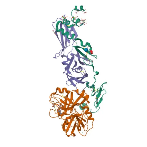
- Structural studies have elucidated the molecular basis of the TF-FVIIa complex, providing insights into the initiation of blood coagulation.
Experimental Guidance and Technical Tips
- The immunogen corresponds to the extracellular domain (amino acids 34–251), suggesting the antibody recognizes the ligand-binding region; confirm reactivity against both membrane-bound and soluble TF in your experimental system.
- For Western blotting, a band of approximately 33 kDa is expected under reducing conditions; include appropriate positive controls (e.g., human placental or lung tissue lysates).
- In immunohistochemistry (IHC-P), antigen retrieval with citrate buffer may enhance staining; optimize primary antibody dilution and detection system for your tissue type.
- For immunofluorescence/ICC, fixation and permeabilization conditions should be optimized to detect both surface and intracellular TF pools.
- For ELISA, consider using paired antibody sets or direct detection after validating linearity and sensitivity with recombinant TF standards.
CamelBio: Your One-Stop Sourcing Bridge
As a premier one-stop IVD raw-material partner, CamelBio streamlines procurement for coagulation research with robust antibodies and supporting reagents. Our supply capabilities extend from validated antibody pairs to bulk monoclonals and rare target sourcing, enabling diagnostic manufacturers to advance projects efficiently from concept to clinic.
REQUEST A QUOTE
Our professional team will reply to you within one business day. Please feel free to contact us!