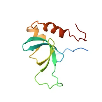
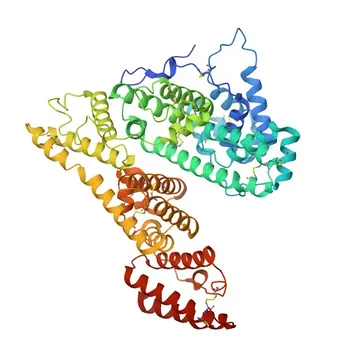
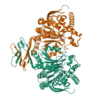
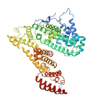
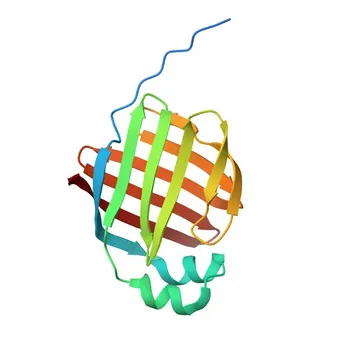
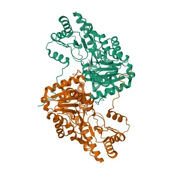
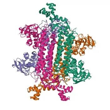
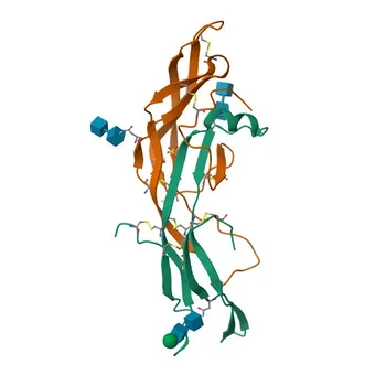
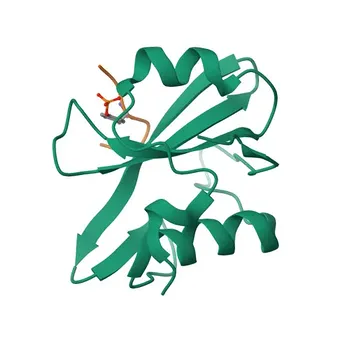
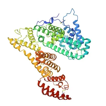
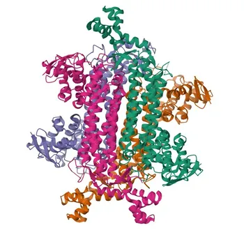
An effective non-invasive hepatic fibrosis assay must measure the dynamic balance between matrix breakdown and inhibition, and the critical serological targets are the matrix metalloproteinases (MMP-2, -3, -9, -13) and the tissue inhibitors of metalloproteinases (TIMP-1, TIMP-2), along with profibrotic cytokines like TGF-β1 and IL-13. These enzymes and their inhibitors directly reflect the ECM turnover driven by activated hepatic stellate cells, and their rising or falling blood concentrations correlate with fibrosis progression or regression, giving assay developers the biochemical backbone for staging liver disease without biopsy.
The core diagnostic opportunity lies in quantifying the net fibrolytic balance: as fibrosis advances, TIMP-1 and TIMP-2 rise sharply, suppressing the collagen-degrading activity of MMPs. Therefore, multiplex panels that measure both the proteases and their endogenous inhibitors provide the most accurate serological snapshot of active ECM remodeling, enabling non-invasive staging and regression monitoring.
The Enzymatic Engine of ECM Remodeling: Matrix Metalloproteinases
When hepatic stellate cells transdifferentiate into myofibroblasts, they reshape the liver’s scaffold by secreting MMPs that can degrade fibrillar collagens. The primary reference identifies four MMPs of particular diagnostic relevance: MMP-2, -3, -9, and -13.
MMP-2 (Gelatinase A): The Constitutive Sentinel
MMP-2 is constitutively expressed and often elevated in early injury. It degrades basement membrane collagen IV and denatured collagens (gelatin), signaling ongoing microarchitectural remodeling.
Its serum levels may rise before significant fibrosis is established, making it a candidate for early detection panels. However, its lack of liver specificity requires careful interpretation.
MMP-3 (Stromelysin-1): The Broad‑Spectrum Activator
MMP-3 activates other pro‑MMPs and cleaves proteoglycans and glycoproteins. Its serum concentration can reflect the extent of matrix turnover beyond just collagen, pulling it into panels that aim to capture the full ECM‑degrading milieu.
It is particularly useful when combined with TIMP-1 measurements because its activity is tightly regulated by the inhibitor, and the resulting ratio can sharpen the diagnostic signal.
MMP-9 (Gelatinase B): The Inflammatory Amplifier
MMP-9 is released largely by Kupffer cells and infiltrating immune cells, linking inflammation to matrix degradation. Its serum elevation often accompanies active hepatitis, so it can help distinguish fibrogenesis driven by ongoing inflammation from burnt‑out cirrhosis.
For assay developers, the challenge is that MMP-9 can spike in systemic inflammation; thus, it must be used in a liver‑focused panel to avoid false positives from extrahepatic sources.
MMP-13 (Collagenase‑3): The True Collagen Executor
MMP-13 directly cleaves the native triple‑helical structure of type I collagen, the dominant fibrillar collagen in cirrhotic liver. It is the human orthologue of rodent MMP-13 (originally defined in the rat) and is markedly downregulated in advanced fibrosis.
Measuring MMP-13 alone rarely suffices, but its decline in parallel with TIMP-1 elevation provides a powerful biochemical fingerprint of impaired collagenolysis that drives scarring.
The Brakes: Tissue Inhibitors of Metalloproteinases
Any diagnostic strategy focused solely on MMPs misses the critical counter‑force: TIMPs. The primary reference emphasizes TIMP-1 and TIMP-2 as the most consequential targets because they progressively rise as fibrosis advances, directly blocking the enzymatic clearance of the scar.
TIMP-1: The Archetypal Fibrosis Marker
TIMP-1 is a 30 kDa glycoprotein whose serum concentration correlates reliably with histological fibrosis stage, from mild portal expansion to established cirrhosis. It inhibits nearly all MMPs but has especial affinity for MMP-9 and MMP-13.
Its progressive increase is the biochemical hallmark of the fibrotic phenotype. Assays that quantify TIMP‑1 alone or in ratio to MMP‑13 or MMP‑9 have repeatedly shown high diagnostic accuracy for advanced fibrosis.
TIMP-2: The Constitutive Regulator with Disease‑Specific Surge
TIMP‑2 is more constitutively expressed but also rises significantly during chronic liver injury, especially in alcohol‑related and cholestatic liver diseases. It preferentially inhibits MMP‑2.
Measuring TIMP‑2 alongside TIMP‑1 can improve the assay’s ability to stage fibrosis across different etiologies, because the TIMP‑2/MMP‑2 ratio captures the local pericellular balance around activated HSCs.
Why the Net Balance Matters More Than Single Analytes
In early fibrosis, MMP activity may predominate, generating a “high‑turnover” state. In advanced cirrhosis, TIMPs overwhelm MMPs, creating a net “low‑turnover” state where scars persist. Therefore, the ratio of TIMP‑1 to MMP‑13 or MMP‑9 is a superior serological index for distinguishing F2 from F4 fibrosis than any single protein concentration.
Regulatory Cytokines as Surrogate Markers of Fibrogenesis
The ECM remodeling process is orchestrated by profibrogenic cytokines, and the primary reference identifies TGF‑β1 and IL‑13 as critical targets for assay developers.
TGF-β1: The Master Fibrogenic Switch
TGF‑β1 is the most potent activator of HSCs into collagen‑secreting myofibroblasts. It also upregulates TIMP‑1 and suppresses MMP‑13 expression, creating a vicious cycle. Its serum levels can be elevated even before detectable matrix changes, making it a candidate for predicting future progression.
However, TGF‑β1 is notoriously latent and platelet‑bound; therefore, immunoassays need rigorous preanalytical standardisation (serum vs. plasma, platelet depletion, activation steps) to yield reproducible results.
IL-13: The Th2‑Linked Fibrosis Promoter
IL‑13 drives fibrosis through a TGF‑β1‑independent pathway, promoting collagen deposition via MMP‑9 dysregulation. Its concentration is especially relevant in hepatitis C and other inflammatory liver diseases.
Including IL‑13 in a multiplex panel helps capture fibrosis driven by immune‑mediated mechanisms that may be missed by TGF‑β1‑centric tests.
Understanding the Trade‑offs: Pitfalls in Assay Development
Despite their diagnostic promise, targeting ECM enzymes and serological markers comes with significant technical and biological challenges that must be addressed to build a robust non‑invasive test.
Preanalytical Variability Can Eclipse the Biological Signal
MMPs and TIMPs are sensitive to collection tube types, clotting, and freeze‑thaw cycles. TIMP‑1 is especially prone to platelet release; even a slight platelet contamination during serum preparation can artefactually elevate TIMP‑1 and distort the MMP/TIMP ratio.
Pitfall: A poorly standardised sample handling protocol can produce TIMP‑1 results that reflect sample quality, not fibrosis stage. Rigorous standardisation of serum vs. plasma, centrifugation speed, and time is non‑negotiable.
Lack of Liver Specificity
All the listed enzymes and inhibitors are expressed in extrahepatic tissues (joints, uterus, tumours). An elevated serum MMP‑9 could indicate hepatic inflammation, rheumatoid arthritis, or malignancy.
Mitigation: No single marker is liver‑specific. Only a carefully normalised panel that considers the pattern of multiple analytes — such as the combination of TIMP‑1, MMP‑13, and TGF‑β1 — can confidently attribute changes to liver pathology.
Dynamic Range and Reverse Zoning
In some patients, MMP‑13 may be undetectable while TIMP‑1 is extremely high, compressing the dynamic range of a ratio‑based index. Diagnostic assay developers must validate the lower limit of quantification (LLOQ) for each analyte and decide whether to use a ratio or a composite score (e.g., logistic regression formula) to accommodate the full spectrum of values.
Making the Right Choice for Your Assay Development Goal
The “best” target depends entirely on the clinical question your assay is designed to answer. Use the following guide to select your core analytes.
- If your primary focus is broad‑spectrum screening for undiagnosed fibrosis: Include TIMP‑1 and MMP‑13 in a ratio‑based panel. This combination captures the net collagenolytic deficit across the majority of etiologies and gives the clearest separation between F0–F2 and F3–F4.
- If your primary focus is early detection of fibrogenesis before structural damage: Add TGF‑β1 and MMP‑2 to the panel. TGF‑β1 flags persistent HSC activation, while MMP‑2 rises during initial basement membrane remodelling, providing lead time before collagen accumulation.
- If your primary focus is monitoring therapy‑induced regression: Measure TIMP‑1, TIMP‑2, and MMP‑9 serially. A decline in the TIMP‑1:MMP‑9 ratio over time has been strongly associated with collagenolytic clearance and histological regression in antiviral and antifibrotic trials.
- If your primary focus is differentiating inflammatory from burnt‑out cirrhosis: Include IL‑13 and MMP‑9. Elevated IL‑13 with a high MMP‑9:TIMP‑1 ratio suggests active, immune‑driven disease, whereas low MMP‑9 with sky‑high TIMP‑1 points to end‑stage low‑turnover cirrhosis.
By selecting the right biochemical levers — the MMPs that degrade, the TIMPs that restrain, and the cytokines that orchestrate — your assay can turn the complex biology of ECM remodeling into a simple, actionable blood test. Focus on the balance, not the individual, and your diagnostic will deliver the clinical value that liver patients urgently need.
Summary Table:
| Target | Biomarker Class | Primary Biological Function | Diagnostic Application & Utility |
|---|---|---|---|
| MMP-2 | Gelatinase | Degrades basement membrane collagen IV | Early-stage matrix remodeling & detection |
| MMP-3 | Stromelysin | Activates pro-MMPs; cleaves proteoglycans | Broad matrix turnover & TIMP-1 ratio modeling |
| MMP-9 | Gelatinase | Driven by immune/Kupffer cell inflammation | Distinguishes active hepatitis from burnt-out cirrhosis |
| MMP-13 | Collagenase | Directly cleaves native type I collagen | Decreases in advanced scarring (impaired collagenolysis) |
| TIMP-1 | Inhibitor | Blocks MMP-9/13; preserves scar tissue | Core marker for staging fibrosis severity (F0–F4) |
| TIMP-2 | Inhibitor | Blocks MMP-2; regulates pericellular matrix | Etiology-specific staging (cholestatic/alcoholic) |
| TGF-β1 | Cytokine | Master activator of stellate cells & TIMP-1 | Early prediction of ongoing fibrogenesis |
| IL-13 | Cytokine | Drives TGF-β1-independent fibrotic pathways | Captures immune-mediated liver fibrosis |
Accelerate Your Liver Fibrosis Assay Development with CamelBio
Developing highly sensitive, reliable multiplex panels for ECM remodeling markers requires top-tier reagents and deep technical expertise. CamelBio provides diagnostic manufacturers, laboratories, and research institutes with one-stop access to high-performance IVD raw materials, technical services, and consulting—covering every stage of your project from concept to clinic.
Whether you need validated antibody pairs for MMP/TIMP ratios, high-purity recombinant proteins, or assay optimization support to overcome preanalytical variability, CamelBio is your trusted partner.
Ready to bring your non-invasive diagnostic test to market? Contact CamelBio Today to request raw material samples or consult with our IVD development team!
Related Products
- Anti-HDGF Monoclonal Antibody for WB, IF-P, ELISA - P51858
- Anti-Alpha-Fetoprotein (AFP) Monoclonal Antibody for WB, IF/ICC, ELISA - P02771
- Alpha-Fetoprotein (AFP) Mouse Monoclonal Antibody for WB, IF/ICC, ELISA - P02771
- Anti-IDH1 Rabbit Monoclonal Antibody for WB, IF/ICC, ELISA - O75874
- Anti-Alpha-Fetoprotein (AFP) Monoclonal Antibody for WB, IF/ICC, ELISA - P02771