Polyclonal Antibodies
Anti-FER Polyclonal Antibody for WB and ELISA - P16591
Item Number : CM0030295
Price varies based on specs and customizations
- Application
- WB, ELISA
- Cross Reactivity
- Human, Mouse, Rat
- Protein Weight
- 95kDa

Shipping:
Contact us to get shipping details Enjoy On-time Dispatch Guarantee.
Why Choose Us
Easy ordering process, quality products, and dedicated support for your business success.
Core Product Specifications and Parameters
| Parameter | Value |
|---|---|
| Product Name | FER Rabbit pAb |
| Remarks/Alias | TYK3; PPP1R74; p94-Fer; FER |
| Species | Human |
| Gene ID (Human) | 2241 |
| Gene ID | 2241 |
| Immunogen | Recombinant fusion protein containing a sequence corresponding to amino acids 200-450 of human FER (NP_005237.2) |
| Source | Rabbit |
| Category | Polyclonal Antibodies |
| Application | WB, ELISA |
| Cross Reactivity | Human, Mouse, Rat |
| SWISS | P16591 |
| Protein Weight | 95kDa |
| Shipping | Ice bag |
Biological Background: FER Function and Localization
- FER (Tyrosine-protein kinase Fer) is an intracellular non-receptor tyrosine kinase belonging to the FES/FPS kinase family; it is also known as TYK3, p94-Fer, and PPP1R74. Isoform 1 is ubiquitously expressed, while isoform 3 is detected in testis and certain carcinomas.
- It acts downstream of growth factor receptors (e.g., EGFR, KIT, PDGFRA/B), mediating signal transduction to promote cell proliferation, differentiation, and survival.
- FER phosphorylates multiple substrates including CTTN, FAK, GAB1, PECAM1, and STAT3, thereby regulating the actin cytoskeleton, microtubule dynamics, lamellipodia formation, and cell adhesion/migration.
- It plays a critical role in leukocyte chemotaxis and transendothelial migration upon LPS stimulation, as well as in mast cell degranulation via the FCER1 pathway.
- In the nervous system, FER contributes to synapse organization, synaptic vesicle trafficking, and neuron-neuron transmission; it is also implicated in neuronal cell death after brain injury.
- FER localizes to the cytoplasm, cytoskeleton, cell cortex, membrane, nucleus, and cell junctions, reflecting its involvement in diverse signaling complexes.
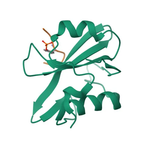
- Key post-translational modifications include phosphorylation, and it contains Src homology 2 (SH2) and protein kinase domains.
Experimental Guidance and Technical Tips
- The immunogen covers amino acids 200-450 of human FER, a region encompassing the SH2 domain and part of the catalytic kinase domain; this design may ensure detection of both full-length and truncated FER isoforms by Western blot.
- For Western blotting, a band at approximately 95 kDa is expected; however, actual observed molecular weight can vary depending on cell type and post-translational modifications.
- The antibody has been validated for WB and ELISA; it demonstrates cross-reactivity with mouse and rat FER, enabling its use across multiple model organisms.
- When performing ELISA, proper antigen coating and blocking conditions should be optimized experimentally to achieve the best signal-to-noise ratio.
- As a polyclonal antibody, lot-to-lot consistency should be monitored; it is advisable to include appropriate positive and negative controls in each assay.
CamelBio: Your One-Stop Sourcing Bridge
CamelBio serves as a one-stop sourcing partner for IVD raw materials, supplying high-quality antibodies, antigens, and ancillary reagents to diagnostic manufacturers worldwide. For research on non-receptor tyrosine kinases like FER, CamelBio facilitates seamless procurement of validated polyclonal and monoclonal antibodies, helping to streamline assay development and validation.
REQUEST A QUOTE
Our professional team will reply to you within one business day. Please feel free to contact us!
Related Articles
qPCR Primer Design: The Small Decisions That Determine Assay Reliability
A practical guide to qPCR primer design, from amplicon length and 3' stability to validation for efficient, reproducible diagnostics.
When Fluorescence Becomes Evidence: Engineering a Competitive ELISA for Trace Molecule Detection
Learn how procedural discipline, blank correction, substrate selection, and reader configuration turn fluorometric ELISA sensitivity into reliable trace detection.
Competitive ELISA vs. GC/MS: Building a Faster, Defensible Strategy for Trace Detection
Learn how competitive ELISA compares with GC/MS for trace detection and how to validate immunoassay kits for reliable laboratory screening.
Choosing the Right High-Throughput Toxicology Platform: Cell-Based Bioassays Versus ELISA
Compare cell-based bioassays and ELISA for toxicology screening, from biological relevance and throughput to validation and technical support.