Prorenin cross-reactivity forces a binary choice in antibody selection: target a conformational epitope exclusive to active renin, or abandon direct mass measurement altogether in favor of an enzyme activity assay.
Because prorenin circulates at concentrations up to 100 times higher than active renin, even a trace of antibody cross-reactivity can swamp the true renin signal. Diagnostic developers must therefore either secure highly specific monoclonal antibodies that bind only the active renin conformation, or adopt enzymatic plasma renin activity (PRA) methods that are inherently immune to prorenin interference.
In plasma, prorenin outnumbers active renin by up to 100:1. An antibody with just 1% cross-reactivity to prorenin will double the measured renin concentration, making clinical reference ranges useless. The only path to a reliable direct plasma renin concentration (PRC) assay is through dual‑antibody sandwich formats that exploit the unique three‑dimensional conformation of the active enzyme’s catalytic cleft—an epitope hidden in prorenin.
The Biology: Why Prorenin Dominates the Problem
The Renin–Prorenin Axis in Circulation
Prorenin is not a rare precursor; it is present at 10 to 100 times the level of active renin in virtually all individuals. Because prorenin contains the entire amino acid sequence of renin, an antibody raised against any linear peptide epitope will almost certainly recognise both forms with equal avidity.
Shared Sequence, Divergent Conformations
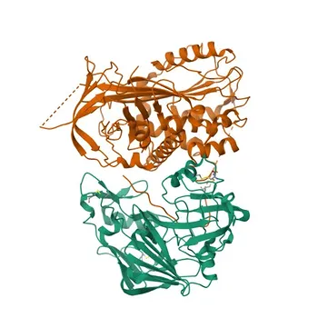
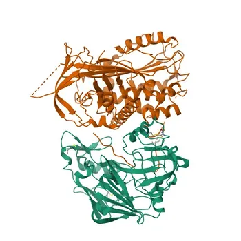
The only reliable immunochemical difference lies in three‑dimensional shape. Prorenin’s prosegment blocks its active site, while active renin exposes a distinctive catalytic cleft. This conformational gap is the unique fingerprint that must be exploited to achieve selectivity.
How Cross-Reactivity Sabotages Assay Accuracy
The Mathematics of Overestimation
Assume prorenin is present at a 100‑fold excess. An antibody with 1% cross-reactivity will generate a prorenin signal equal to the entire active renin signal, doubling the result. At 5% cross-reactivity, the reported value becomes six times the true concentration—a catastrophic error that destroys any dose‑response relationship.
Clinical Fallout of a False‑High Renin Result
Overstated renin mass can lead to misdiagnosis of hyperreninemic hypertension, unnecessary renal artery imaging, and misguided therapy. A direct assay that cannot distinguish active hormone from its inactive precursor is clinically dangerous.
Strategic Antibody Selection for Direct PRC Assays
Conformational Epitopes: The Key to Excluding Prorenin
The core strategy is to use monoclonal antibodies that bind exclusively to the three‑dimensional active site of renin. Because prorenin’s catalytic cleft is sterically buried, these antibodies see only active renin, eliminating the bulk of the interfering signal at the first binding event.
Dual‑Antibody Sandwich Formats
A single conformational antibody reduces cross‑reactivity. Pairing two such antibodies—a capture and a detection antibody both targeted to the active cleft—creates a double‑lock that multiplies selectivity. This is the foundation of modern immunoradiometric (IRMA) and immunochemiluminometric (ICL) direct renin assays.
Rigorous Screening and Validation
Candidate antibody pairs must be tested against isolated prorenin and clinical samples with known high prorenin levels. Spiking experiments, comparison with enzymatic PRA as a reference method, and calibration against international renin standards (e.g., WHO IRP 68/356) are all compulsory steps to confirm true prorenin‑blind performance.
The Alternative Pathway: Enzymatic Plasma Renin Activity
PRA Avoids the Cross-Reactivity Trap Entirely
Plasma renin activity assays measure the generation of angiotensin I, a reaction catalysed only by active renin. Prorenin is enzymatically silent under standard handling conditions, so PRA is naturally free of prorenin interference and serves as the reference method when antibody specificity cannot be guaranteed.
PRA vs. PRC: Different Measures, Different Clinical Utility
PRA reflects the functional activity of renin within the patient’s own plasma milieu, but it is sensitive to substrate concentration and sample preparation. Direct PRC assays measure absolute mass and can be easier to automate, yet their fidelity depends entirely on the antibody’s ability to reject prorenin.
Understanding the Trade-offs and Pitfalls
The Price of Extreme Specificity
Conformation‑specific antibodies are rare and often exhibit lower affinity than those binding linear epitopes. This can constrain assay sensitivity, require more complex signal‑amplification steps, and increase manufacturing cost—all factors that must be weighed against the clinical imperative of accuracy.
Incidental Prorenin Activation in the Lab
Prorenin can be cryo‑activated or acid‑activated during sample handling, forcibly exposing the catalytic cleft. Even a well‑chosen antibody will then bind activated prorenin and produce false‑high results. Rigid pre‑analytical protocols are essential to prevent this hidden source of overestimation.
Harmonisation Challenges Across Platforms
Because different manufacturers use antibody pairs targeting different active‑renin epitopes, direct PRC assays are not analytically interchangeable. This mirrors the known variability seen in insulin and natriuretic peptide immunoassays, meaning that clinical decision limits cannot simply be transferred from one kit to another without extensive cross‑validation.
Making the Right Choice for Your Assay Development Goal
- If your primary focus is absolute freedom from prorenin interference: Develop or source two high‑affinity monoclonal antibodies directed against non‑overlapping conformational epitopes inside the active renin cleft, and validate them in a robust sandwich IRMA/ICL format.
- If your primary focus is rapid development and proven robustness: Design an enzymatic PRA assay that eliminates prorenin cross‑reactivity by its biochemical design, even if it demands more hands‑on laboratory steps.
- If your primary focus is high‑throughput, automated testing: Push for a direct PRC sandwich assay, but be prepared for exhaustive antibody screening and enforce strict pre‑analytical handling to forestall inadvertent prorenin activation.
- If your primary focus is cross‑platform harmonisation: Anchor your assay to an international reference material, openly characterise your epitope specificity, and recognise that full inter‑assay agreement will require industry‑wide collaboration.
The true measure of a direct renin immunoassay is not how effectively it captures renin, but how completely it ignores prorenin. Make that discrimination your design imperative, and your assay will deliver the clinical accuracy patients depend on.
Summary Table:
| Strategy / Format | Target Epitope | Primary Advantage | Key Challenge / Risk |
|---|---|---|---|
| Conformational Sandwich PRC | Active site 3D catalytic cleft | High-throughput automation & high specificity | Rare high-affinity antibodies; risk of sample cryo-activation |
| Enzymatic PRA Assay | Enzymatic generation of Ang I | Inherently immune to prorenin interference | Variable substrate concentration; labor-intensive workflow |
| Linear Epitope Antibodies | Shared peptide sequences | High binding affinity & lower cost | Severe cross-reactivity (up to 100:1 excess); false-high results |
Developing high-specificity assays requires precise antibody pairing to eliminate prorenin cross-reactivity. CamelBio provides IVD diagnostic manufacturers, labs, and research institutes with one-stop access to high-quality IVD raw materials, technical services, and expert consulting—covering every stage from concept to clinic. Ready to optimize your assay performance? Contact us today to collaborate with our technical team!
Related Products
- Anti-Renin Monoclonal Antibody for WB, IF/ICC, ELISA - P00797
- Anti-Renin Rabbit Polyclonal Antibody for WB, IHC-P, ELISA - P00797
- Anti-Nephrin Polyclonal Antibody for WB, IF, IHC, ELISA - Q9QZS7
- Anti-Nephrin Rabbit Monoclonal Antibody for WB, IF-P, IHC-P, ELISA - Q9QZS7
- Anti-RING1B/RNF2 Monoclonal Antibody for WB, IHC-P, ELISA - Q99496