Uncompromising sensitivity and batch-to-batch consistency define the competitive edge of modern diagnostic immunoassays.
In vitro display technologies like ribosome display and mRNA display directly answer these demands by enabling the screening of antibody libraries of unprecedented size and integrating built-in affinity maturation. They let you find rare, high-affinity binders—often with dissociation constants in the picomolar range—and then continuously evolve their properties to meet the strict detection limits of ELISA, CLIA, and lateral flow platforms.
The core advantage is twofold. First, eliminating cellular transformation bottlenecks unlocks library diversities of 1012–1015 variants, vastly increasing the odds of finding high-affinity hits for difficult diagnostic targets. Second, the cell-free format allows you to embed error-prone PCR directly between selection rounds, enabling rapid, iterative optimization of affinity, specificity, and thermal stability without ever re-cloning into a host.
Breaking Through the Diversity Ceiling
Why Cell-Free Equals Limitless
Traditional phage display and hybridoma libraries are capped by bacterial transformation efficiency or the natural B-cell repertoire—usually around 1010–1011 clones.
Ribosome and mRNA display operate entirely in vitro. No living cells are needed to express or amplify the library, so the only limit is the amount of DNA you can handle in a tube. This routinely yields 1012 to 1015 unique variants, a 100‑ to 10,000‑fold increase.
The Impact on Diagnostic Sensitivity
Larger libraries directly translate into a higher probability of discovering ultra‑high‑affinity clones with dissociation constants (Kd) in the low nanomolar to picomolar range.
For diagnostic platforms like chemiluminescent immunoassays (CLIA) or high‑sensitivity ELISAs, this means you can detect low‑abundance biomarkers that would otherwise fall below the noise floor. The difference between a 10 nM binder and a 50 pM binder often determines whether an assay is clinically useful.
Evolution in a Tube: Continuous Affinity Maturation
Built‑in Mutagenesis via Error‑Prone PCR
The hallmark of ribosome and mRNA display is the physical linkage of the antibody fragment to its encoding mRNA. After target binding, the mRNA of enriched variants is recovered and reverse‑transcribed.
Because you amplify that genetic material with non‑proofreading polymerases or use error‑prone PCR conditions, you can intentionally introduce random mutations across the variable domains. These mutations can hit not just CDRs but also framework residues, opening up new affinity‑enhancing solutions that are fully acceptable for diagnostic raw materials.
Faster Optimization Without Re‑cloning
The entire cycle—binding, washing, elution, RT‑PCR with mutagenesis, and re‑transcription—stays in one seamless tube‑based workflow.
You can perform 3–5 rounds of affinity maturation in a few weeks, driving a mediocre binder from micromolar to picomolar affinity without ever pausing to transform, clone, or sort cells. This dramatically shortens the development timeline compared to hybridoma or phage display methods that require subcloning between rounds.
Understanding the Trade‑offs
From Display to Detection: The Reformatting Challenge
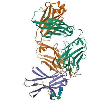
Most in vitro display selections yield antibody fragments such as scFv or Fab.
For use in commercial IVD kits, these often need to be reformatted into full‑length IgGs or engineered fusion proteins (e.g., scFv‑Fc, Fab‑biotin). While well‑established, reformatting can occasionally alter affinity or introduce soluble‑expression bottlenecks in mammalian or E. coli systems. Early pilot expression and functional testing in the final assay format is essential.
Soluble Expression and Aggregation Risk
Not every high‑affinity clone that shines in a cell‑free translation system will fold correctly when produced at scale. Framework mutations introduced during affinity maturation may improve paratope geometry but destabilize the overall immunoglobulin fold.
Diagnostic developers must screen lead candidates for monomeric purity, thermal stability, and low‑aggregation tendency before committing to production.
Managing Background in Ultra‑Large Libraries
With 1014 variants, you also have 1014 opportunities for non‑specific stickiness. Rigorous counter‑selection against sample matrix components (e.g., albumin, interfering proteins) and stringent washing conditions are mandatory.
Otherwise, you risk isolating high‑affinity plastic‑ or matrix‑binders that look great on selection but fail in real serum or plasma samples.
Making the Right Choice for Your Diagnostic Program
The decision to use ribosome or mRNA display should align with your assay’s most critical technical hurdle.
- If your primary focus is ultra‑sensitive detection of a low‑abundance biomarker: Start with a large naïve library (>1012 clones) to pull out pM‑affinity binders, then apply error‑prone PCR maturation to push the detection limit even lower.
- If your target is a highly conserved, toxic, or non‑immunogenic antigen: Leverage the cell‑free format’s ability to bypass animal immune tolerance; screen directly against the native antigen without animal immunization.
- If batch‑to‑batch consistency and scalable supply are your top priorities: Once a lead is identified, freeze the DNA sequence and transition to recombinant production in E. coli or CHO cells. This guarantees a permanent, reproducible supply that far surpasses hybridoma‑derived reagents.
- If time‑to‑market is critical: Combine in vitro display with an established reformatting pipeline; you can go from target to validated scFv or Fab reagents in as little as 8 weeks, then rapidly integrate them into your IVD platform.
By fusing unlimited library size with programmable evolution, in vitro display platforms deliver the affinity, specificity, and reproducibility that diagnostic developers need to stay ahead of ever‑more‑sensitive detection standards.
Summary Table:
| Key Feature / Benefit | Traditional Platforms (Phage/Hybridoma) | In Vitro Display (Ribosome/mRNA) | Diagnostic Impact |
|---|---|---|---|
| Library Diversity | 10⁹–10¹¹ clones (cell-constrained) | 10¹²–10¹⁵ variants (cell-free) | Higher probability of capturing rare, picomolar-affinity binders |
| Affinity Maturation | Requires re-cloning & transformation | Built-in via tube-based error-prone PCR | Continuous optimization of affinity, specificity, and stability |
| Development Speed | Months of iterative cell work | Target to validated hit in ~8 weeks | Dramatically shortened timeline to market for IVD assays |
| Target Flexibility | Restricted by cellular toxicity & immunogenicity | Cell-free, direct screening against native target | Enables screening against toxic, non-immunogenic, or conserved targets |
| Supply Reproducibility | Risk of cell drift or loss | Recombinant sequence defined | Guaranteed batch-to-batch consistency for commercial assay kits |
Accelerate Your Assay Development with CamelBio
High-sensitivity, reliable diagnostic immunoassays depend on superior binder quality and batch-to-batch consistency. CamelBio provides diagnostic manufacturers, labs, and research institutes with one-stop access to premium IVD raw materials, recombinant antibody technical services, and expert consulting—covering every stage from concept to clinic.
Ready to enhance your ELISA, CLIA, or lateral flow platform sensitivity? Contact us today to collaborate with our IVD experts and streamline your antibody selection pipeline.
Related Products
- Anti-T-bet/Tbx21 Rabbit Recombinant Monoclonal Antibody for WB - Q9UL17
- Anti-JAM-A/CD321/F11R Rabbit Monoclonal Antibody for WB, IF/ICC, ELISA - Q9Y624
- Anti-REST/NRSF Rabbit Polyclonal Antibody for WB, ELISA - Q13127
- Anti-RAP1A + RAP1B Monoclonal Antibody for WB, IF/ICC, ELISA - P61224 / P62834
- Anti-REST/NRSF Rabbit Monoclonal Antibody for WB, IP, ELISA - Q13127
People Also Ask
- What are the challenges in scaling up recombinant proteins & mAbs? Scale-Up Guide
- Why Use Recombinant Monoclonal Antibodies for Pathogen Detection? Key Benefits for IVD Developers
- What factors determine whether an IVD antibody uses bacterial vs. mammalian expression?
- What are the differences between batch culture and semipermeable-membrane mAb production?
- How do parasitic immune evasion mechanisms impact IVD raw material selection? Optimize Serological Assay Design