The Tube Looks Quiet Until the Signal Moves
A real-time PCR assay can appear perfectly controlled during setup.
The primers are present. The polymerase is active. The target sequence is theoretically specific. The instrument is ready to record fluorescence.
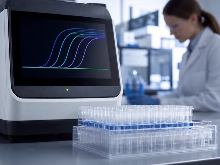
Then, near the end of the run, the no-template control begins to rise.
Nothing was added to the tube. No pathogen was present. Yet the curve resembles amplification.
This is the diagnostic danger of primer-dimers: a reaction can produce evidence without producing the intended target.
Primer-dimers are short, non-specific double-stranded products formed when primers transiently bind to one another and are extended by DNA polymerase. In research, they may be an inconvenience. In diagnostics, they can become a system-level failure involving chemistry, interpretation, and clinical risk.
The Molecular Event Behind a False Signal
The 3′ End Is Where the Problem Begins
Primer-dimers are not random accidents caused simply by having two primers in the same tube.
The critical trigger is complementarity at the primers' 3′ termini.
When the forward and reverse primers share several complementary bases at their 3′ ends, they can briefly anneal to one another. If the alignment is stable enough, the polymerase treats one primer as a template for the other.
A short duplex becomes an extendable substrate.
Once extended, that initial product can serve as a template in later cycles. The artifact then follows the same exponential logic as a legitimate amplicon.
| Molecular feature | What happens |
|---|---|
| 3′ complementarity | Primers anneal to each other |
| Polymerase access | One primer extends from the other |
| First dimer product | A short double-stranded artifact is created |
| Subsequent cycles | The artifact is amplified exponentially |
Three or more complementary bases at the 3′ termini can be sufficient to create this chain of events, depending on sequence context and reaction conditions.
High primer concentration and elevated GC content can increase the opportunity for interaction, but they are not the fundamental cause by themselves. Without a vulnerable 3′ alignment, those factors do not reliably create a primer-dimer.
The First Risk May Occur Before Cycling
The thermal cycler is not necessarily where the problem starts.
During reaction setup, the mixture may remain at room temperature for several minutes. Conventional polymerases can retain partial activity during this period. A transient primer-primer interaction that would otherwise disappear may be extended before the initial denaturation step.
That early extension matters.
The reaction has effectively received a head start on the wrong template. Subsequent cycles do not need to create the artifact again; they only need to copy it.
This is why the physical workflow of an assay matters as much as its final cycling protocol. Time at ambient temperature, order of reagent addition, and enzyme chemistry can all influence the final signal.
How a Small Artifact Changes Assay Performance
Intercalating Dyes Cannot Tell the Difference
SYBR Green and related intercalating dyes respond to double-stranded DNA.
They do not know whether that DNA is the intended amplicon or a short primer-dimer. If the artifact reaches sufficient concentration, it produces fluorescence and may generate an apparent amplification curve.
The instrument reports fluorescence. The analyst supplies the biological meaning.
That gap is where false positives enter.
A melt-curve analysis can sometimes distinguish a primer-dimer from the intended product because the artifact often has a lower melting temperature. But this is a corrective interpretation step, not a substitute for clean assay design. A diagnostic workflow that depends on subjective curve interpretation carries avoidable uncertainty.
Reagent Depletion Raises the Ct Value
Primer-dimers compete with the intended target for the same limited resources:
- Primers
- dNTPs
- DNA polymerase
- Magnesium ions
- Reaction time within the exponential phase
Even when the dimer does not produce a visible false-positive signal, it can consume enough reagents to weaken target amplification.
The result may be a higher Ct value, reduced amplification efficiency, and a narrower dynamic range. The effect is especially important near the limit of detection, where only a small number of target copies are available.
At high target concentrations, the intended amplicon may out-compete the artifact. At low target concentrations, the artifact may gain a disproportionate advantage.
Multiplex Reactions Add Another Layer of Risk
In multiplex qPCR, several primer pairs occupy the same reaction environment.
This creates additional possibilities:
- Self-dimers within one primer
- Homodimers between primers of the same orientation
- Cross-dimers between different assay targets
- Competition for polymerase and dNTPs
- Fluorescence interpretation errors across channels
A primer pair that performs well in a singleplex reaction can behave differently when introduced into a multiplex panel. The assay is no longer a collection of independent reactions. It is a network of interacting sequences sharing one chemical system.
That system must be evaluated as a whole.
The Conditions That Make Primer-Dimers More Likely
Primer Concentration
Primer concentrations above approximately 0.5 µM can increase the frequency of random collisions and stabilize weak interactions.
The highest concentration is not automatically the most sensitive choice. More primer can increase the raw rate of target initiation, but it can also increase background amplification.
A practical starting point is approximately 0.2 µM of each primer, followed by controlled titration based on Ct value, efficiency, and non-specific signal.
Magnesium Concentration
Magnesium ions stabilize DNA duplexes and support polymerase activity.
Too little magnesium can impair target amplification. Too much can stabilize imperfect primer interactions and promote non-specific products.
The correct concentration is therefore a balance, not a fixed universal value. Magnesium should be reduced to the lowest level that preserves efficient and reproducible target amplification.
Secondary Structures
A primer can interact with itself before it encounters the target.
Hairpins and self-dimers consume reagent capacity and may create structures that seed larger non-specific products. These risks can be easy to overlook when the primary design review focuses only on target binding.
A complete design assessment should examine:
- Forward-primer self-complementarity
- Reverse-primer self-complementarity
- Forward/reverse cross-complementarity
- 3′ terminal interactions
- Hairpin stability
- Interactions between all primer pairs in a multiplex panel
A Layered Strategy for Preventing Primer-Dimers
No single intervention solves every primer-dimer problem. Reliable diagnostic development uses several layers of control.
1. Design Primers for the Reaction You Actually Need
Primer design should begin with the intended clinical and analytical use, not just the target sequence.
A robust initial screen typically includes:
- Primer length between 15 and 30 bases
- Forward and reverse Tm values within approximately 2°C
- Moderate GC content
- Minimal 3′ cross-complementarity
- Minimal self-dimer and hairpin formation
- No avoidable 3′ terminal thymidine
- No unnecessary GC clamp at the 3′ end
- Amplicon placement that preserves relevant variant coverage
Design software can identify candidate interactions, but software output still requires experimental validation. The most thermodynamically attractive primer pair may not be the most clinically useful one if it compromises variant detection or creates a difficult amplicon.
2. Use a Hot-Start Polymerase
Hot-start polymerases remain inactive, or substantially less active, during reaction setup.
They become fully active only after the initial denaturation or activation step. This prevents transient primer pairs formed at room temperature from being extended into stable artifacts.
Hot-start chemistry is particularly valuable when:
- Reactions are assembled manually
- Many samples are prepared simultaneously
- The setup period is extended
- Primer concentrations must remain relatively high
- The assay targets low-copy nucleic acids
- Multiplex specificity is critical
Antibody-based and chemically modified hot-start enzymes are common approaches. Their value is not limited to reducing primer-dimers; they also improve control over non-specific amplification during the most vulnerable stage of the workflow.
3. Separate Amplification from Signal Generation
Sequence-specific probes add a second recognition requirement.
A hydrolysis probe, such as a TaqMan-style probe, must bind the intended amplicon before a fluorescent signal is generated. A primer-dimer may still exist, but if it lacks the probe-binding sequence, it remains largely invisible to the detection system.
This is a powerful design principle:
Amplification can be imperfect without making every imperfection visible as a positive result.
Probe-based detection increases reagent cost and development complexity. It also requires careful attention to probe design, fluorophore compatibility, and multiplex channel performance. For diagnostic assays where false positives carry substantial consequences, that added specificity is often worth the investment.
4. Build Controls into Every Run
Controls should be part of the assay architecture, not an afterthought.
At minimum, include no-template controls to monitor reagent contamination and background dimerization. For more demanding workflows, also consider:
- Positive controls for target recovery
- Extraction controls for process monitoring
- Internal amplification controls for inhibition
- Serial dilutions for efficiency and linearity
- Replicate low-copy samples near the detection limit
- Melt-curve or product analysis where intercalating dyes are used
A no-template control that remains negative is not proof that no primer-dimers exist. It is evidence that they did not cross the method's reporting threshold under those conditions.
Matching the Mitigation Strategy to the Risk
Different assays require different levels of protection.
| Primary assay objective | Preferred strategy | Main trade-off |
|---|---|---|
| Maximum clinical specificity | Hot-start polymerase plus sequence-specific probe | Higher per-reaction cost and development effort |
| Cost-efficient screening | Rigorous primer design, optimized Mg²⁺ and primer levels, routine NTCs | Greater dependence on melt-curve and amplification analysis |
| Detection of low-copy targets | Hot-start chemistry, low but effective primer concentration, dilution validation | Excessive primer reduction may weaken signal |
| Multiplex detection | Inter-pair dimer screening plus probes for each target | More complex optimization and fluorescence management |
| High-throughput manufacturing | Stable reagent formulation and controlled setup conditions | May require additional validation of storage and workflow robustness |
The correct decision depends on the consequence of error.
If a false positive can trigger unnecessary treatment or compromise clinical trust, reducing reagent cost is rarely the only meaningful optimization target. If the assay is designed for broad screening, a dye-based system may remain appropriate, provided that its limitations are understood and controlled.
Engineering is often the discipline of choosing which failure modes are acceptable.
Validation Should Follow the Assay's Weakest Point
A primer-dimer control strategy is incomplete until it is tested under stress.
Validation should examine more than a clean high-copy sample. It should include conditions that expose competition and non-specific amplification:
- Low-copy target concentrations
- No-template controls
- Long setup intervals at room temperature
- Multiple reagent lots
- Different operators
- Planned primer concentration ranges
- Magnesium concentration ranges
- Potentially interfering sample matrices
- Multiplex combinations at realistic target ratios
Track more than the presence or absence of a curve.
Useful performance indicators include:
- Ct shift across primer concentrations
- Amplification efficiency
- Linearity across serial dilutions
- Limit of detection
- False-positive rate in NTCs
- Melt-curve profile for dye-based assays
- Reproducibility near the decision threshold
- Cross-reactivity with non-target sequences
A primer-dimer problem often reveals itself as instability before it reveals itself as a dramatic false positive. Small Ct shifts, increasing replicate variance, or unexplained late curves are early warnings that the reaction is losing control.
The Economics of Specificity

Every mitigation strategy has a cost.
Hot-start polymerases can increase per-reaction expense and may require longer activation times. Probe-based systems require additional oligonucleotides, fluorescent labels, and design work. Lower primer concentrations may reduce background but also reduce fluorescence intensity or compromise low-copy detection.
Primer redesign can create a different kind of cost. A new amplicon may avoid dimerization but perform poorly against emerging variants or degraded clinical samples.
The right question is not, “Which chemistry is cheapest?”
It is:
What is the total cost of an incorrect result, a repeated run, a failed validation batch, or a diagnostic product that clinicians do not trust?
For manufacturers, this calculation extends beyond the individual reaction. It includes raw-material consistency, scale-up behavior, technical transfer, lot release, and the ability to maintain performance from concept to clinic.
Where CamelBio Supports Assay Development

Primer-dimers are a molecular problem, but solving them often requires decisions across several development stages.
CamelBio provides diagnostic manufacturers, clinical laboratories, and research institutes with one-stop access to IVD raw materials, technical services, and consulting. This support spans the path from assay concept through clinical implementation.
Development teams may need to:
- Select a hot-start enzyme with the right activity profile
- Compare probe-based and intercalating-dye chemistries
- Optimize primer, magnesium, and enzyme concentrations
- Investigate non-specific amplification
- Validate low-copy performance
- Improve reagent consistency across lots
- Prepare an assay for manufacturing or clinical use
These decisions are connected. A change in polymerase can alter the optimal magnesium concentration. A change in primer concentration can shift the limit of detection. A switch to probe detection can solve false fluorescence while introducing new multiplex design requirements.
The useful partner is therefore not only a supplier of isolated components. It is a source of compatible raw materials, technical knowledge, and development guidance that can be applied across the entire assay system.
Final Perspective
A primer-dimer begins as a small event: a few complementary bases meet at the wrong end of two oligonucleotides.
The consequences can become much larger.
The artifact can consume reagents, distort Ct values, generate false fluorescence, obscure low-copy targets, and create cross-talk in multiplex panels. More importantly, it can turn a technically plausible assay into a clinically unreliable one.
The strongest qPCR assays treat primer-dimer prevention as a system-level design task. They combine careful sequence selection, hot-start chemistry, appropriate detection architecture, optimized reaction conditions, and controls that expose failure before patients do.
For support with raw materials, qPCR chemistry, assay optimization, and the path from concept to clinic, Contact Our Experts.
Related Products
- Anti-PKC zeta Polyclonal Antibody for WB, IF/ICC, ELISA - Q05513
- Monoclonal Mouse IgG1 Isotype Control Antibody (Conjugate) for Flow Cytometry - Mouse IgG 1 isotype control
- N6-methyladenosine (m6A) Rabbit Polyclonal Antibody for meRIP, Dot Blot, ELISA, and Nucleotide Array
- Anti-APITD1 Rabbit pAb - Q8N2Z9
- Anti-Catalase Polyclonal Antibody for WB, IHC-P, IF/ICC, ELISA - P04040
Related Articles
- From Matrix Noise to Measurable Signal: A Practical Extraction Strategy for ELISA Testing
- Matrix Interference in ELISA Development: Designing Accuracy Into Complex-Sample Assays
- When the Dye Sees Everything: A Systematic Strategy for Cleaner, More Reliable qPCR
- qPCR Primer Design: The Small Decisions That Determine Assay Reliability
- Why Direct Hapten Coupling Changes the Economics of Small-Molecule Biosensors