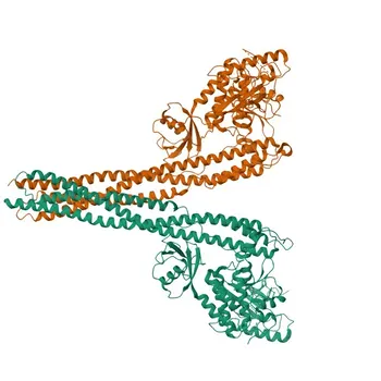
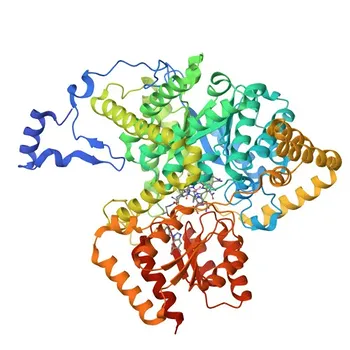
A single enzymatic cleavage. The fluorogenic substrate 4‑methylumbelliferone‑β‑D‑galactoside (4‑MUG) is hydrolyzed by the enzyme β‑galactosidase to release the highly fluorescent molecule 4‑methylumbelliferone (4‑MU). The reaction is then quenched with an alkaline stop solution—typically sodium carbonate—which simultaneously halts enzyme activity and deprotonates the 4‑MU, maximizing and stabilizing its fluorescence for precise quantitative measurement.
The core mechanism is a β‑galactosidase‑catalyzed hydrolysis of a glycosidic bond, followed by an alkaline shift that converts 4‑MU into its strongly fluorescent dianionic form. This simple, two‑step reaction provides the foundation for high signal‑to‑noise ratios, low detection limits, and broad dynamic ranges in fluorogenic immunoassays.
The Two‑Step Chemical Mechanism
Enzymatic Hydrolysis of the Glycosidic Bond
The signal generation begins when a β‑galactosidase‑labeled detection antibody binds to its target. Upon addition of 4‑MUG, the enzyme’s active site specifically recognizes the β‑D‑galactoside moiety.
The reaction cleaves the glycosidic bond between the galactose sugar and the 4‑methylumbelliferone aglycone. This hydrolysis liberates free 4‑MU in solution.
The cleavage is highly specific and proceeds at a rate proportional to the amount of enzyme present. Because β‑galactosidase has a high turnover number, a single enzyme molecule generates many 4‑MU molecules, providing inherent signal amplification.
Alkaline Termination and Fluorophore Activation
Once sufficient product has formed, an alkaline stop solution is added. Common formulations use sodium carbonate to raise the pH above 10.
At neutral pH, 4‑MU exists primarily in its protonated, weakly fluorescent phenol form. The alkaline environment deprotonates the 7‑hydroxyl group, converting it to the phenolate (dianionic) species. This form exhibits a quantum yield that is orders of magnitude higher.
The high pH also denatures the β‑galactosidase, instantly stopping further substrate turnover. The resulting solution is then stable, and the fluorescence can be measured at the optimal wavelengths—excitation around 365 nm and emission around 448 nm.
Why This Mechanism Delivers Superior Signal Generation
High Enzymatic Turnover
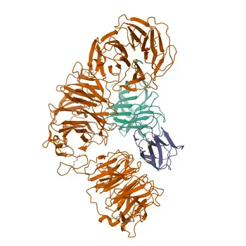
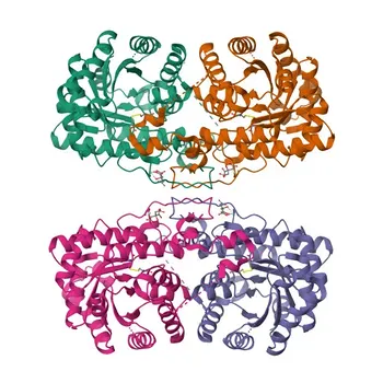
β‑galactosidase is a robust enzyme with a large molecular structure, allowing it to process many substrate molecules per second. Even at low conjugate concentrations, the accumulated 4‑MU signal becomes easily detectable.
This enzymatic amplification directly translates to higher analytical sensitivity. Immunoassays built on this chemistry can reliably measure analyte concentrations that are orders of magnitude lower than the actual enzyme label present.
Low Background and Wide Dynamic Range
The 4‑MUG substrate itself is essentially non‑fluorescent until cleaved. In its intact form, the glycosidic bond quenches the fluorescence of the 4‑MU core.
This chemical quietness keeps intrinsic background extremely low. Combined with the pH‑activated jump in fluorescence after stopping, the system achieves outstanding signal‑to‑noise ratios.
A cleaner baseline means the assay can discriminate minute signal changes from near‑zero noise. For diagnostic kit developers, this empowers a broader dynamic range and more confident cut‑off determinations.
Understanding the Trade‑offs
Substrate Stability and Spontaneous Hydrolysis
4‑MUG is stable under storage and during the incubation period at neutral pH. However, it can undergo slow, non‑enzymatic hydrolysis if inadvertently exposed to alkaline conditions before the stop step.
Premature exposure generates fluorescent background and compromises limit‑of‑detection claims. Keeping the substrate solution buffered at neutral to slightly acidic pH and minimizing light exposure are critical for maintaining lot‑to‑lot consistency.
Process Timing and Hands‑On Steps
The two‑step workflow—incubation followed by alkaline stop—introduces a manual timing element. Variation in the incubation time or delay before adding the stop solution can alter the final signal.
Automated liquid handling systems mitigate this, but in manual kits, it demands precise operator technique. Some users may prefer one‑step substrates that develop continuously, but the MUG/β‑gal approach often rewards that extra pipetting step with superior signal quality.
Making the Right Choice for Your Diagnostic Assay
Whether you are formulating a new kit or optimizing an existing one, the decision to use the 4‑MUG/β‑galactosidase system should align with your performance priorities and workflow constraints.
- If your primary focus is achieving the lowest possible detection limit: The low intrinsic background and high enzyme turnover of this system make it an excellent candidate, provided you can control incubation timing precisely.
- If your primary focus is a robust, user‑friendly workflow: Consider automating the stop step or evaluating whether an alkaline phosphatase/4‑MUP system may offer a simpler single‑step kinetics read; however, be prepared for a potential trade‑in background characteristics.
- If your primary focus is high signal stability for batch processing: The alkaline stop simultaneously halts the reaction and locks in the fluorescence, allowing plates to be read minutes or even hours later without signal drift.
The hydrolysis mechanism behind 4‑MUG is ultimately an elegantly simple way to convert a silent reporter into a brilliant, quantifiable signal—giving you the control you need to build assays that truly push performance boundaries.
Summary Table:
| Reaction Step / Parameter | Key Chemical Mechanism | Analytical Benefit |
|---|---|---|
| 1. Enzymatic Hydrolysis | β-galactosidase cleaves the glycosidic bond of 4-MUG to release 4-MU | High signal amplification via enzyme turnover |
| 2. Alkaline Shift | Stop solution (pH > 10) deprotonates 4-MU to its phenolate form | Maximizes fluorescence yield and locks signal |
| Excitation / Emission | 365 nm (Excitation) / 448 nm (Emission) | Optimal spectral separation with minimal overlap |
| Substrate Quietness | Intact 4-MUG is non-fluorescent at neutral pH | Ultra-low background and broad dynamic range |
Ready to optimize your fluorescent immunoassay development? CamelBio provides diagnostic manufacturers, labs, and research institutes with one-stop access to high-quality IVD raw materials, technical services, and expert consulting—covering every stage from concept to clinic. Contact us today to enhance your assay sensitivity and lot-to-lot consistency!
Related Products
- PKC delta Rabbit Polyclonal Antibody for WB, IHC-P, IF/ICC, ELISA - Q05655
- Anti-BTLA Rabbit Polyclonal Antibody for WB, ELISA - Q7Z6A9
- Anti-PKC zeta Polyclonal Antibody for WB, IF/ICC, ELISA - Q05513
- Anti-FER Polyclonal Antibody for WB and ELISA - P16591
- Anti-BDNF Monoclonal Antibody (Mouse) for WB, ELISA - P23560
People Also Ask
- What performance specs must cortisol kits satisfy for cosyntropin testing? Key Analytical Limits
- How do latex particle physical and chemical properties influence the performance of light scattering immunoassays?
- Why are surfactants required in nitrocellulose membranes? Optimize Antibody Compatibility in Assays
- How do substituent modifications on acridinium carboxamides impact kinetics & stability? Master IVD Label Tuning
- Why are N-sulfonylacridinium-9-carboxamides preferred over acridinium phenyl esters in CLIAs? Key IVD Advantages