To answer that directly: Diagnostic manufacturers must engineer Galectin-3 immunoassays to overcome two primary physiological confounders—renal function interference and high baseline biological variation—while achieving the analytical rigor required for long-term prognosis. This means the assay cannot simply provide a number; it must provide a stable, reproducible number across a comorbid patient population where renal function is often compromised.
The core challenge is separating the cardiac signal from physiological noise. A successful assay is not the one with the highest raw sensitivity, but the one that is clinically validated against renal impairment and age-related changes, backed by rigid calibration standards and precisely defined reference intervals to provide a reliable, actionable prognosis.
The Two-Front War: Physiological Confounders
The prognostic power of Galectin-3 in heart failure is uniquely vulnerable to a patient’s overall metabolic state. A poorly designed assay will reflect kidney function more than cardiac fibrosis, leading to clinical mistrust.
The Dominant Challenge: Renal Cross-Interference
The single most critical biological variable you must account for is renal clearance. Galectin-3 accumulates systemically when kidney function is impaired, which is a direct analytical trap.
Baseline levels are elevated simply due to reduced glomerular filtration. The assay result must be interpreted as a composite marker of fibro-inflammatory burden and renal clearance, not just a pure cardiac scar signal. If your assay cannot differentiate these contributions, its prognostic specificity in a heart failure cohort—where renal insufficiency is common—plummets.
The fixation on a single numeric decision threshold is a strategic mistake. You must establish and validate renal-function-stratified reference intervals. A level of 18 ng/mL in a patient with normal GFR carries a very different weight than the same value in a patient with Stage 4 Chronic Kidney Disease. The assay design, and more importantly the clinical validation data, must actively guide this interpretation.
The Baseline Instability Factor
Beyond the kidneys, physiological baseline variation creates a low-grade analytical buzz. Galectin-3 is a pro-miscuous, pro-inflammatory lectin that rises with age, general cardiovascular stress, and metabolic disturbances.
Establishing a single "normal range" is analytically treacherous. The high biological variation demands that your reference population studies be impeccably stratified by age and comorbidity profile. Statistical outliers in a "healthy" population are often individuals with undiagnosed cardiovascular disease or declining renal function.
Prognostic assays must be optimized for stability at the median, not just sensitivity at the ceiling. While high-sensitive troponin focuses on the 99th percentile, Galectin-3’s chronic prognostic value relies on tracking a rise from a patient-specific baseline within the 3.8-21.0 ng/mL range. This requires exceptional inter-run precision and long-term calibration consistency to detect a medically meaningful delta over months of testing.
Architecting an Assay for Physiological Reality
Translating these physiological constraints into a viable IVD product requires a methodological approach rooted in rigid analytical design controls. Your reagent selection and validation protocol must directly counteract the known biological instability.
Forging a Reliable Quantitative Signal
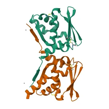
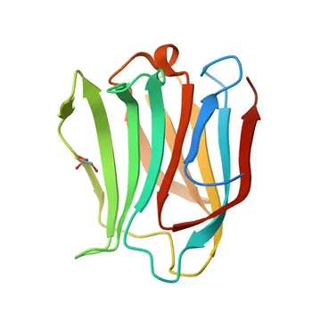
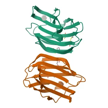
The assay’s physical chemistry must be tuned for the Galectin-3 molecule's specific behavior. Galectin-3 is a β-galactoside-binding lectin, a characteristic you can exploit for solid-phase capture or signal generation, but which also requires careful matrix management.
Focus on rigorous bound-free separation to mitigate heterophilic noise. General immunoassay development rules are critical here. Choosing a high-capacity solid-phase capture system and perfecting the bound-free separation step ensures that the signal represents true analyte concentration, cutting through the non-specific protein noise common in complex comorbidity samples.
The Non-Negotiable Validated Performance Package
Regulatory approval hinges on demonstrating bioanalytical mastery. Because of the subjective nature of the prognosis, your validation data must be stronger than for an acute diagnostic.
High compliance in precision, selectivity, and stability is non-negotiable. You must demonstrate low %CV at the clinically relevant decision levels, prove assay selectivity against other galectin family members and common interfering substances, and guarantee long-term reagent stability. Supply chain confidence, built on high-grade IVD raw materials, ensures that batch-to-batch consistency doesn’t become the variable that breaks the prognostic trend over years of patient monitoring.
Defining Truth: The Quest for the Reference Standard
The most profound analytical hurdle is the absence of a pure gold standard for "free, biologically active Galectin-3." Standardization must be defined intra-assay.
The reference interval is your product’s intellectual core. It must be established using a rigorous, biologically plausible calibration standard and confirmed across a demographically diverse cohort that honestly represents the renal function spectrum of a real-world heart failure population. A reference interval built only on healthy 30-year-olds is worthless for a 75-year-old HF patient.
Understanding the Trade-offs
Building a trusted assay involves making deliberate, transparent technical sacrifices to satisfy the clinical need.
Analytical Sensitivity vs. Biological Specificity: Chasing femtogram-level sensitivity is a wasted effort. The biological noise floor from renal and inflammatory cross-talk is high. Resources are better spent on maximizing precision within the clinically significant 3-20 ng/mL window and developing robust matrix management protocols.
Generalization vs. Renally Stratified Cut-offs: It is analytically simpler to publish one universal decision threshold. However, this creates clinical confusion and risks product misapplication. The trusted, scientifically rigorous path is to invest in the validation necessary to provide adjusted nomograms or percentage-predicted models that account for estimated GFR. This turns a physiological weakness into a diagnostic feature.
Making the Right Choice for Your Development Goal
The path you prioritize in assay development will define your product's clinical positioning and commercial success.
- If your primary focus is long-term prognostic monitoring: Design the assay for maximum longitudinal stability and inter-run precision. Invest heavily in calibrator lot traceability so a value measured in year one is directly comparable to a value measured in year three.
- If your primary focus is risk stratification at a single point-of-care: Validate your single-point cut-offs against a population that accurately reflects the full renal function continuum of your target market. The clinical utility hinges on demonstrating the cut-off’s positive predictive value is not compromised by a high prevalence of renal insufficiency.
- If your primary focus is entering a regulated new market: Prioritize your design control documentation for selectivity and the establishment of your reference range. A defense of your reagent raw material traceability is the foundational argument for regulatory approval to achieve clearance as a Class II or similar quantitative immunoassay device.
Your goal is to build an assay that clinicians trust not because it's new, but because it honestly reflects the complex biology of the patients it serves, enabling a confident prognosis even in the face of physiological ambiguity.
Summary Table:
| Factor / Challenge | Clinical & Analytical Impact | Recommended Assay Design Strategy |
|---|---|---|
| Renal Cross-Interference | Reduced GFR elevates baseline Gal-3, causing false prognostic signals. | Validate GFR-stratified reference intervals and renally adjusted nomograms. |
| Biological Variation | Systemic inflammatory noise and age-related drift complicate cut-offs. | Focus on precision within the 3–20 ng/mL decision window over extreme sensitivity. |
| Matrix Noise & Non-Specific Binding | Lectin-binding properties risk non-specific interferences in serum. | Optimize high-capacity solid-phase capture and strict bound-free separation protocols. |
| Long-Term Monitoring Requirements | Patient monitoring occurs over years; lot-to-lot shift ruins trend accuracy. | Ensure lot traceability and rely on robust, highly consistent IVD raw materials. |
Developing a robust Galectin-3 assay requires high-purity antibodies, reliable calibrators, and proven analytical expertise. CamelBio provides diagnostic manufacturers, labs, and research institutes with one-stop access to premium IVD raw materials, technical services, and consulting—covering every stage from concept to clinic.
Overcome physiological confounders and ensure superior batch-to-batch consistency for your cardiac biomarker assays—contact our IVD experts today!
Related Products
- Anti-Galectin 3/LGALS3 Rabbit Monoclonal Antibody for WB, IHC-P, IF/ICC, ELISA - P17931
- [KO Validated] Anti-Galectin-3/LGALS3 Rabbit Polyclonal Antibody for WB, IF/ICC, ELISA - P17931
- Galectin 3/LGALS3 Rabbit Monoclonal Antibody for WB, IHC-P, IP, ELISA - P17931
- Anti-Galectin 3/LGALS3 Rabbit Monoclonal Antibody for WB, IF/ICC, IP, FC, ELISA - P17931
- Anti-Human Galectin-3 Monoclonal Antibody for Flow Cytometry (Intracellular) - P17931

![[KO Validated] Anti-Galectin-3/LGALS3 Rabbit Polyclonal Antibody for WB, IF/ICC, ELISA - P17931](https://image.camelbiological.com/images/products/6658/main_image_6a63e48ad76336.06622480-small.webp)