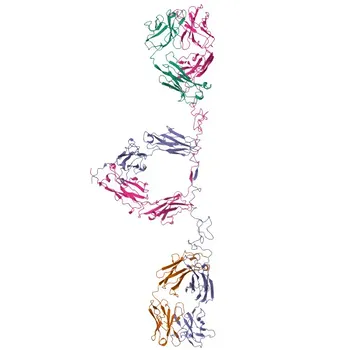
Acute hemolytic transfusion reactions (AHTRs) are driven by two distinct antibody-mediated pathways.
IgM isoagglutinins (anti-A, anti-B) trigger complement activation and rapid intravascular hemolysis, while IgG antibodies against non-ABO antigens cause extravascular clearance via the reticuloendothelial system. For IVD developers, these mechanisms directly dictate the need for assays that can sensitively detect both antibody classes—using high-avidity monoclonal antibodies for direct agglutination and anti-human globulin (AHG) reagents to reveal non-agglutinating IgG alloantibodies before transfusion.
The core immunological challenge of pre-transfusion screening is that clinically dangerous antibodies can be either IgM or IgG, and they often exist at low titers. Therefore, blood group typing reagents must provide immediate, high-avidity agglutination of carbohydrate antigens, while antibody screening assays must amplify the signal from weakly bound IgG using a validated AHG test—both of which rely on precisely characterized raw materials that mirror the underlying type II hypersensitivity mechanism.
The Immunological Mechanisms of AHTR
To design an assay that prevents hemolysis, you must first understand exactly how the immune system destroys transfused red blood cells.
Complement-Dependent Intravascular Hemolysis by IgM
Naturally occurring IgM anti-A and anti-B antibodies are the primary culprits in ABO-mismatched transfusions.
These large, pentameric antibodies efficiently fix complement, generating the membrane attack complex that lyses donor red cells directly in the bloodstream.
This reaction is rapid, catastrophic, and requires no “sensitization” period—because these isoagglutinins are pre-formed from early childhood.
The assay equivalent must, therefore, be able to produce immediate, visible agglutination without incubation, using direct hemagglutination with high-titer IgM monoclonal reagents.
Fc-Mediated Extravascular Clearance by IgG
Non-ABO alloantibodies (e.g., anti-Rh, anti-Kell, anti-Duffy) are usually IgG, which does not fix complement efficiently in the circulation.
Instead, the antibody-coated red cells are recognized by Fcγ receptors on macrophages in the spleen and liver, leading to phagocytosis and extravascular destruction.
Because IgG rarely causes direct agglutination in saline, the antibody screening test must bridge this gap.
This is achieved by adding an AHG reagent (anti-human IgG) that cross-links sensitized cells, converting invisible binding into detectable agglutination.
Translating Mechanism to Assay Requirements
The immunological mechanism of AHTR is not just academic; it defines every critical performance parameter in a pre-transfusion IVD.
Blood Group Typing: Direct Agglutination Demands High-Avidity IgM Reagents
ABO typing must work immediately at room temperature, as an emergency mismatch would be lethal.
This means the monoclonal anti-A and anti-B antibodies must possess exceptionally high avidity for the terminal carbohydrate epitopes—GalNAc for A, galactose for B.
The reagents must be validated against panels of defined RBCs to confirm no cross-reactivity with the O group (which lacks both terminal sugars).
Any low-affinity binding can cause weak or false-negative reactions, directly risking a mistransfusion.
Antibody Screening: Revealing “Invisible” IgG with the Indirect Antiglobulin Test
The screen for unexpected antibodies cannot rely on direct agglutination; most clinically significant alloantibodies are IgG and will not agglutinate erythrocytes in a low-ionic medium alone.
Therefore, the test must incorporate the indirect antiglobulin test (IAT), where washed RBCs are first sensitized with patient serum, then incubated with an anti-human IgG AHG reagent.
The sensitivity of this AHG reagent is the single most critical variable for detecting low-titer, high-affinity antibodies.
It must contain a broad specificity anti-IgG that recognizes all subclasses, while avoiding prozone effects that can yield false negatives at very high antibody concentrations.
The Red Cell Panel: A Defined Antigenic Landscape
You cannot screen for antibodies without a comprehensive, well-characterized panel of reagent red cells.
Each cell in the panel must be phenotyped for clinically significant antigens (Rh, Kell, Duffy, Kidd, etc.), so that a specific pattern of reactivity can identify an antibody’s target.
This panel must balance antigen representation and stability—rare antigens may be absent, but common ones that cause severe hemolysis must be reliably present.
The cells themselves must be preserved in a way that maintains membrane integrity and antigen expression, as degradation can abolish epitopes and generate false negatives.
Critical Raw Materials and Their Roles
Pairing the right raw materials with the correct immunological target is the linchpin of a successful IVD assay.
Monoclonal Antibodies for Blood Group Antigens
For forward typing, monoclonal IgM anti-A and anti-B are the gold standard because they directly agglutinate native RBCs without requiring AHG.
For reverse typing (testing patient plasma against known A₁ and B cells), the same high-avidity properties apply to the panel cells, not the reagent.
In antigen phenotyping of donor cells (e.g., RhD typing), high-affinity monoclonal IgM anti-D reagents must not cause false positives with weak D variants, requiring careful epitope mapping and blending of clones to cover all clinically relevant D epitopes.
Anti-Human Globulin (AHG) Reagents for the IAT
The AHG reagent for IgG detection is a polyspecific or IgG-specific antibody raised against human IgG heavy chains.
It must be free of heterophile antibodies that could agglutinate uncoated cells, and its dilution must be optimized so that the bridging signal is strong without causing non‑specific aggregation.
Additionally, some protocols include anti-C3d in the AHG reagent to detect complement-fixing IgM or IgG antibodies that may have bound C3 fragments to the cell surface, widening the net for rare AHTR-causing antibodies.
Control Sera and Antigen Standards
Every test run needs positive and negative control sera containing known antibody specificities (e.g., anti-D, anti-K) to verify that the entire detection system—from red cell panel to AHG—is functioning.
For antigen controls, recombinant or purified blood group substances (such as synthetic A/B trisaccharides) can be used to confirm antibody specificity in inhibition studies, ensuring that the monoclonal antibody reagent is not cross-reacting with structurally similar epitopes.
Understanding the Trade-offs
No assay design can maximize every parameter simultaneously; acknowledging the compromises is essential for building a trustworthy product.
Sensitivity vs. Specificity
Pushing the AHG test to detect femtomolar IgG levels risks false-positive reactions from clinically insignificant antibodies or cold autoantibodies.
False positives lead to unnecessary transfusion delays and extensive workups, eroding clinical confidence in the test.
Conversely, a less sensitive AHG reagent may miss a weak but hemolytic anti-Jk(a) antibody, exposing the patient to a delayed but real risk of extravascular destruction.
Manufacturers must strike a balance by validating sensitivity against a panel of clinically significant antibodies with known minimum titers.
Monoclonal Antibody Avidity and Cross-reactivity
Extremely high-avidity anti-A IgM will agglutinate Ax or Ael subgroups, which have very few A antigens; this can be clinically correct but may confuse typing algorithms if not clearly defined.
Cross-reactivity with bacterial polysaccharides or other blood group systems must be excluded by testing against rare null phenotype red cells and using inhibition assays with purified antigens.
Panel Cell Representation and Shelf-Life
A panel covering all rare antigens is prohibitively expensive and may have limited stability.
Most screening panels focus on antigens responsible for ≥0.1% of clinically significant AHTRs, accepting that some rare antibodies (e.g., anti-Vel) will only be detected at the crossmatch stage.
Preservation media can alter membrane rigidity, potentially exposing cryptic antigens or masking ones critical for robust agglutination.
Every new lot must be cross-validated with a reference method to ensure no shift in reactivity pattern.
Making the Right Choice for Your IVD Development Goal
Your assay’s configuration must align with the specific immunological vulnerabilities you aim to detect. Here is how to navigate the key decision points.
- If your primary focus is ABO front‑typing accuracy: Select monoclonal IgM reagents with extreme avidity and specificity for the terminal carbohydrate epitopes, and validate them against a comprehensive panel of weak subgroups and null phenotypes to eliminate dangerous false negatives.
- If your primary focus is detecting clinically significant IgG alloantibodies: Invest in a high‑sensitive AHG reagent that covers all IgG subclasses and include a complement (anti‑C3d) component, while defining a clear reactivity threshold to minimize serological “noise” from cold autoantibodies.
- If your primary focus is a screening panel for donor centers: Engineer the cell panel to maximize antigen representation for high‑incidence, clinically significant markers (Rh, Kell, Duffy, Kidd, Ss), and implement rigorous lot‑to‑lot monitoring to catch any loss of weak antigen expression before it causes a missed alloantibody.
- If your primary focus is a low‑cost point‑of‑care test: Optimize a direct agglutination only (IgM‑driven) assay with lyophilized monoclonal blood grouping reagents, clearly labeling that it is not a substitute for a full antibody screen using an AHG‑based IAT.
The immunological pathway of AHTR is unforgiving, but it is also fully understood. By mapping each step—antigen structure, antibody class, and effector mechanism—to a specific raw material and assay parameter, you can build a pre‑transfusion screening system that turns this mechanistic clarity into unmatched patient safety.
Summary Table:
| Pathway / Mechanism | Target Antibody & Antigen | Required Assay Strategy | Critical Raw Materials |
|---|---|---|---|
| Intravascular Hemolysis | IgM (Anti-A, Anti-B) on ABO antigens | Direct hemagglutination (Immediate at RT) | High-avidity monoclonal IgM antibodies |
| Extravascular Clearance | IgG (Anti-Rh, Kell, Duffy) on non-ABO antigens | Indirect Antiglobulin Test (IAT) bridging | Broad-spectrum anti-human IgG (AHG) reagents |
| Alloantibody Screening | Low-titer IgG / IgM unexpected antibodies | Defined RBC antigen landscape | Phenotyped reagent red cell panels & control sera |
Accelerate your blood group typing and antibody screening assay development with CamelBio. As a trusted partner in immunohematology, CamelBio provides diagnostic manufacturers, labs, and research institutes with one-stop access to premium IVD raw materials, technical services, and consulting—covering every stage from concept to clinic.
Looking to optimize high-avidity monoclonal reagents or validated AHG formulations for your next-generation assay? Contact us today to discuss your project!
Related Products
- MHC class I (HLA-A/B) Rabbit mAb - P30443/P01889/P01891/P01892/P13746/P16188/P30450
- Anti-CA3 Polyclonal Antibody for WB, IF/ICC, ELISA - P07451
- Anti-ABI3 Rabbit Monoclonal Antibody for WB, IHC-P, ELISA - Q9P2A4
- Rabbit anti-Human/Monkey HLA-DR mAb for FC - P01903
- Anti-Emerin/EMD Polyclonal Antibody for WB, IHC-P, IF/ICC, ELISA - P50402
People Also Ask
- How does antigen selection impact HIV-1/HIV-2 differentiation immunoassays? Optimize Sensitivity & Specificity
- What factors determine whether an IVD antibody uses bacterial vs. mammalian expression?
- What are the principles and key reagent considerations when developing bead-based luminescent proximity assay kits?
- How do co-existing structural hemoglobin variants impact HbA2 quantification? Methodologies & Solutions