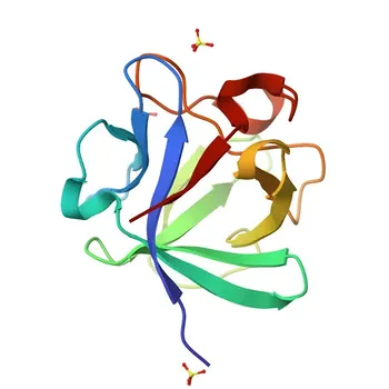
The master phosphate regulator. Fibroblast Growth Factor 23 (FGF-23) is a bone‑derived hormone that directly controls phosphate balance by driving renal phosphate excretion and shutting down active vitamin D (1,25(OH)₂D₃) synthesis. In chronic kidney disease, FGF‑23 rises early to defend against phosphate overload, often long before parathyroid hormone (PTH) or calcitriol levels change. This unique position at the apex of the bone‑kidney‑parathyroid axis makes FGF‑23 an indispensable biomarker for immunoassay panels targeting bone and mineral metabolism disorders in renal patients—it reveals the disease process when conventional markers are still silent.
FGF‑23 is the first‑line phosphate sensor and phosphaturic effector. Its early elevation in kidney disease provides a diagnostic window that PTH and 1,25(OH)₂D₃ alone cannot offer. Pairing FGF‑23 with these traditional markers creates a holistic axis view, enabling earlier detection of CKD‑mineral and bone disorder, better differentiation of phosphate‑wasting syndromes, and more precise monitoring of therapeutic interventions.
The Physiological Role of FGF‑23 in Mineral Homeostasis
FGF‑23 is not simply one more hormone in the calcium‑phosphate cascade—it is the central phosphate‑regulating peptide that integrates signals from bone to kidney.
Where FGF‑23 Comes From and What Triggers It
FGF‑23 is a 32 kDa peptide secreted primarily by osteocytes in bone.
Its production is stimulated by sustained elevations in plasma phosphate and dietary phosphate intake.
This bone‑kidney axis ensures that when phosphate levels climb, the skeleton signals the kidneys to clear the excess.
How FGF‑23 Lowers Phosphate: The Renal Proximal Tubule
FGF‑23 acts in the kidney by binding to a klotho‑FGFR1 receptor complex on the basolateral membrane of proximal tubular cells.
This triggers internalization and lysosomal degradation of the apical sodium‑phosphate cotransporters NPT2a and NPT2c, which normally reclaim filtered phosphate from the tubular lumen.
The result is a rapid, powerful increase in urinary phosphate excretion—a direct phosphaturic effect that lowers serum phosphate.
FGF‑23’s Double Action on Vitamin D Metabolism
Beyond phosphate excretion, FGF‑23 suppresses the renal 1α‑hydroxylase enzyme (CYP27B1), the catalyst that converts 25‑hydroxyvitamin D to active 1,25(OH)₂D₃.
Simultaneously, it may enhance the catabolic 24‑hydroxylase pathway.
This dual suppression starves the system of active vitamin D, which in turn reduces intestinal phosphate and calcium absorption—a secondary mechanism to lower phosphate load.
A Brake on Parathyroid Hormone
FGF‑23 also directly suppresses PTH synthesis and secretion from the parathyroid glands under normal physiology.
This creates a classic endocrine negative‑feedback loop: elevated phosphate triggers FGF‑23, which lowers phosphate and dampens PTH.
However, in renal disease this brake fails, as we will explore.
Why FGF‑23 Becomes Critical in Renal Disease
In chronic kidney disease (CKD), the phosphate‑regulating system becomes chaotic. FGF‑23 is the first responder—and its failure defines the transition to CKD‑mineral and bone disorder (CKD‑MBD).
The Earliest Biomarker of Phosphate Disturbance
As nephrons are lost, the kidney’s capacity to excrete phosphate declines.
FGF‑23 rises exponentially in CKD stage 2–3, well before any measurable change in serum phosphate, PTH, or 1,25(OH)₂D₃.
This compensatory spike maintains normophosphatemia for a long time—but at a cost.
FGF‑23 Resistance and the Shattered Feedback Loop
In advanced CKD, the parathyroid glands become resistant to FGF‑23’s suppressive effects.
Klotho expression in the parathyroids and kidneys drops, and the FGF‑23 receptor complex loses sensitivity.
The result: FGF‑23 levels skyrocket without effectively lowering PTH, contributing to secondary hyperparathyroidism and renal osteodystrophy.
The Link to Calcification and Cardiovascular Risk
High FGF‑23 is now recognized as a potent independent risk factor for left ventricular hypertrophy (LVH), vascular calcification, and mortality in CKD.
FGF‑23 can directly induce cardiac myocyte hypertrophy via klotho‑independent pathways when concentrations are extremely high.
This transforms FGF‑23 from a mere phosphate regulator into a clinically actionable marker of systemic toxicity.
The FGF‑23–PTH–Calcitriol Axis: A Triad of Interlocking Signals
No single marker tells the full story. The physiological dance between FGF‑23, PTH, and 1,25(OH)₂D₃ is why an integrated panel is essential.
How the Three Hormones Regulate Each Other
- PTH stimulates renal 1α‑hydroxylase, increasing 1,25(OH)₂D₃ and thus bone resorption and phosphate release.
- 1,25(OH)₂D₃ stimulates FGF‑23 production by osteocytes, while also suppressing PTH via negative feedback.
- FGF‑23 suppresses both 1,25(OH)₂D₃ synthesis and PTH secretion, closing the loop.
In health, this triad maintains stable mineral levels.
In renal disease, the dissociation of these relationships—high FGF‑23, low 1,25(OH)₂D₃, high PTH—becomes the hallmark of CKD‑MBD.
Disentangling Renal Phosphate Wasting From Parathyroid Disease
An isolated PTH measurement cannot distinguish between primary hyperparathyroidism and renal tubular phosphate‑wasting disorders like autosomal dominant hypophosphatemic rickets (ADHR) or X‑linked hypophosphatemia (XLH).
In XLH, FGF‑23 is inappropriately high (due to a processing defect), causing phosphate wasting with low 1,25(OH)₂D₃ despite normal renal function and often normal PTH.
Only by including FGF‑23 can you correctly classify the disorder and avoid unnecessary parathyroid work‑ups.
Understanding the Trade‑offs: Assay Design and Clinical Interpretation
From an immunoassay developer’s perspective, targeting FGF‑23 demands careful strategic decisions. The molecule’s biology creates distinct design challenges.
Intact FGF‑23 vs. C‑Terminal Fragments: An Assay Dichotomy
Circulating FGF‑23 exists as both the biologically active intact hormone (iFGF23) and inactive C‑terminal fragments (cFGF23).
- An intact assay uses a sandwich pair against both N‑ and C‑terminal epitopes, capturing only the full‑length hormone. It reflects acute biological activity.
- A C‑terminal assay measures total FGF‑23 (intact plus fragments) and can indicate cumulative production. However, results may not directly correlate with phosphate‑wasting activity.
Each format provides a different clinical perspective, and reference intervals are not interchangeable.
Assay developers must validate which population they aim to measure and clearly communicate the test’s interpretive limitations.
Pre‑analytical Stability and Matrix Effects
Intact FGF‑23 is heat‑ and protease‑sensitive; it can deteriorate rapidly at room temperature, leading to falsely low values.
C‑terminal fragments are more stable but may overestimate bioactive hormone in conditions where cleavage is impaired.
Strict specimen handling protocols (EDTA plasma, cold processing, prompt freezing) are mandatory, and kit developers must establish robust stability data.
Renal Function‑Dependent Reference Ranges
Unlike PTH, for which established CKD staging ranges exist, FGF‑23 reference values shift dramatically with glomerular filtration rate.
A “normal” FGF‑23 in a healthy individual may be pathologic in advanced CKD, and vice versa.
Developing stage‑specific or eGFR‑adjusted interpretative ranges is a critical need for clinical adoption.
The Cost of Panel Complexity
Adding FGF‑23 to a PTH‑calcitriol panel increases reagent costs, multiplexing complexity, and regulatory burden.
However, the diagnostic value—earlier intervention, differentiation of hypophosphatemic syndromes, cardiovascular risk stratification—often justifies the investment.
The key is to design the panel so that each marker adds non‑overlapping information, avoiding costly redundancy.
Making the Right Choice for Your Immunoassay Panel
Your decision to include FGF‑23 must be driven by the specific clinical need you aim to address. Here is how to align your assay design with your target application:
- If your primary focus is early CKD‑MBD screening: Include an intact FGF‑23 assay alongside PTH. FGF‑23 elevation precedes PTH rise and provides the earliest signal for nephrologist intervention.
- If your primary focus is differential diagnosis of hypophosphatemia: Use an intact or C‑terminal FGF‑23 assay combined with 1,25(OH)₂D₃. High FGF‑23 with low calcitriol points to phosphate‑wasting syndromes like XLH or ADHR; low FGF‑23 suggests malnutrition or non‑renal losses.
- If your primary focus is cardiovascular risk stratification in dialysis patients: Consider measuring C‑terminal FGF‑23 (or intact, with strict pre‑analytical controls) as an independent mortality predictor, beyond PTH and traditional mineral markers.
- If your primary focus is therapeutic monitoring of phosphate binders or vitamin D analogs: A panel that tracks FGF‑23, PTH, and 1,25(OH)₂D₃ together reveals whether treatment is restoring axis harmony or simply shifting the pathology.
FGF‑23 is no longer a research curiosity—it is the missing voice in the mineral metabolism conversation, and incorporating it into your immunoassay panel transforms a fragmented snapshot into a comprehensive physiological narrative.
Summary Table:
| Biomarker | Primary Physiological Role | Early CKD (Stage 2–3) Dynamics | Essential Immunoassay Design Consideration |
|---|---|---|---|
| FGF-23 | Enhances renal phosphate excretion & suppresses 1,25(OH)₂D₃ synthesis | Rises exponentially before serum phosphate or PTH change | Choose Intact (active) vs. C-terminal (total); strict sample cold chain needed |
| PTH | Stimulates bone resorption & renal 1α-hydroxylase activity | Elevates secondary to FGF-23 resistance & falling calcitriol | Key for staging secondary hyperparathyroidism & renal osteodystrophy |
| 1,25(OH)₂D₃ | Promotes intestinal calcium & phosphate absorption | Suppressed early by rising FGF-23 levels | Crucial for assessing endocrine feedback integrity & active vitamin D deficiency |
Accelerate Your Immunoassay Panel Development with CamelBio
Developing high-performance diagnostic assays for complex bone and mineral metabolism markers requires robust raw materials and proven technical expertise. CamelBio provides diagnostic manufacturers, labs, and research institutes with one-stop access to IVD raw materials, technical services, and consulting—covering every stage from concept to clinic.
Whether you need high-affinity antibodies for intact FGF-23, specialized antigens, or custom assay optimization support, our team is ready to streamline your commercialization path.
Contact us today to discover how CamelBio can support your IVD development goals!
Related Products
- Anti-FGF23 Rabbit Monoclonal Antibody for WB, IF/ICC, ELISA - Q9GZV9
- FGF23 Rabbit Polyclonal Antibody for WB, IF/ICC, ELISA - Q9GZV9
- Anti-FGFR2 Polyclonal Antibody for WB, IF/ICC, ELISA - P21802
- Anti-FGF1 Monoclonal Antibody for WB, IF/ICC, ELISA - P05230
- Anti-FGFR2 (KO Validated) Monoclonal Antibody for WB, ELISA - P21802
![[KD Validated] Anti-FGFR3 Polyclonal Antibody for WB, FC, ELISA - P22607](https://image.camelbiological.com/images/products/9806/main_image_6a641d04ca6008.75267463-small.webp)