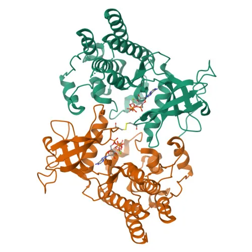
Monoclonal Antibodies
Anti-FGFR2 (KO Validated) Monoclonal Antibody for WB, ELISA - P21802
Item Number : CM0002436
Price varies based on specs and customizations
- Application
- WB, ELISA
- Cross Reactivity
- Human, Mouse, Rat
- Protein Weight
- 92kDa

Shipping:
Contact us to get shipping details Enjoy On-time Dispatch Guarantee.
Why Choose Us
Easy ordering process, quality products, and dedicated support for your business success.
Core Product Specifications and Parameters
| Parameter | Value |
|---|---|
| Product Name | [KO Validated] FGFR2 Rabbit mAb |
| Remarks/Alias | BEK; JWS; BBDS; CEK3; CFD1; ECT1; KGFR; TK14; TK25; BFR-1; CD332; K-SAM; R2 |
| Species | Human |
| GeneID (Human) | 2263 |
| GeneID | 2263 |
| Immunogen | Synthetic Peptide: A synthetic peptide corresponding to a sequence within amino acids 1-100 of human FGFR2 (P21802). |
| Source | Rabbit |
| Category | Monoclonal Antibodies |
| Application | WB, ELISA |
| Cross Reactivity | Human, Mouse, Rat |
| SWISS | P21802 |
| Protein Weight | 92kDa |
| Shipping | Ice bag |
Biological Background: FGFR2 Function and Localization
- FGFR2 (Fibroblast growth factor receptor 2) is a receptor tyrosine kinase that binds fibroblast growth factors, regulating cell proliferation, differentiation, migration, apoptosis, and embryonic development.
- Alternative splicing generates isoforms such as BEK (epithelial isoform) and KGFR (keratinocyte growth factor receptor), which have distinct ligand-binding specificities.
- Core signaling pathways involve phosphorylation of PLCG1, FRS2, and PAK4, leading to activation of RAS-MAPK, AKT1, and STAT1 cascades.
- FGFR2 is essential for osteoblast differentiation and skeletal development; it promotes proliferation in immature osteoblasts but induces apoptosis in mature osteoblasts.
- Subcellular localization includes the cell membrane, Golgi apparatus, cytoplasmic vesicles, and secreted forms; internalization and degradation regulate signaling.
- Post-translational modifications include glycosylation, disulfide bond formation, phosphorylation, and ubiquitination (Ubl conjugation), affecting stability and signaling.
- Genetic mutations in FGFR2 are linked to craniosynostosis, ectodermal dysplasia, lacrimo-auriculo-dento-digital syndrome, and other skeletal dysplasias.
- FGFR2 is classified under keywords including alternative splicing, proto-oncogene, and tyrosine-protein kinase, highlighting its roles in cancer and development.
Experimental Guidance and Technical Tips
- This antibody is KO validated, meaning it has been confirmed to specifically recognize FGFR2 in knockout cell lines; use appropriate positive and negative controls in Western blot and ELISA experiments.
- The immunogen corresponds to the N-terminal region (aa 1-100) of human FGFR2; consider that this region may be conserved across isoforms and species, supporting the reported cross-reactivity with human, mouse, and rat.
- For Western blot, expect a band around 92 kDa; validate in the relevant sample system and consider potential splice variants or degradation products.
- For ELISA, optimize coating and detection conditions using recombinant FGFR2 protein or cell lysates; validate in the relevant matrix.
- When investigating FGFR2 signaling, pair this antibody with phosphorylation-specific antibodies or receptor activation assays to assess functional modulation.
CamelBio: Your One-Stop Sourcing Bridge
CamelBio provides diagnostic manufacturers, laboratories, and research institutes with integrated access to IVD raw materials, including validated antibody pairs, optimized monoclonal/polyclonal antibodies, and ancillary reagents. Our FGFR2 antibody product supports research in cell signaling, skeletal development, and oncology, and we can source bulk quantities or rare target raw materials to streamline your assay development pipeline. With end-to-end technical support, CamelBio accelerates your journey from concept to clinical application.
REQUEST A QUOTE
Our professional team will reply to you within one business day. Please feel free to contact us!