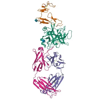
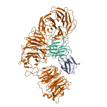
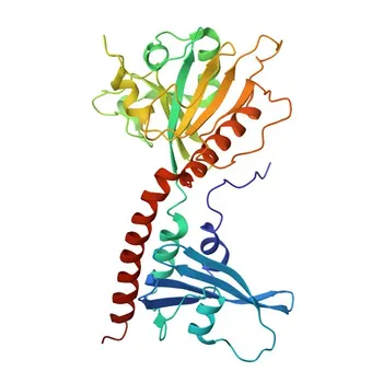
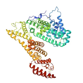
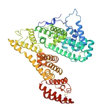
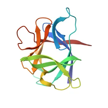
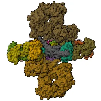
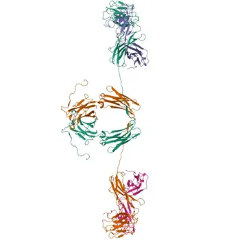
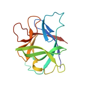
The single most critical distinction is the presence of a non-amino acid partner. Simple proteins consist exclusively of amino acids linked by peptide bonds, while conjugated proteins are built from a protein backbone (the apoprotein) to which a non-protein prosthetic group is permanently attached – such as a carbohydrate, lipid, or metal ion. This structural divide is not academic; it fundamentally dictates how diagnostic antibodies recognize the target and therefore guides the selection of every raw material, from calibrators to detection reagents.
The core challenge of immunoassay raw material development is deciding whether the epitope an antibody must bind to is born from the protein’s pure amino acid sequence or from its marriage to a prosthetic group. Neglecting the prosthetic group can replace a high-affinity binding event with a silent assay, because even a perfect recombinant apoprotein will fail to present the three-dimensional features that a native conjugated protein creates.
The Fundamental Structural Divide
The difference between simple and conjugated proteins lies at the molecular level and cascades upward into practical assay design.
Simple Proteins: Pure Polypeptide Chains
Simple proteins are linear or folded chains of amino acids and nothing else. Examples like serum albumin or insulin contain no built-in non-protein modifications. All structural information comes from the sequence and its resulting three-dimensional fold. That means every potential epitope is coded directly by the gene.
Conjugated Proteins: A Protein Core with Non-Protein Partners
Conjugated proteins are hybrid molecules. A protein part (apoprotein) is covalently or tightly associated with a distinct chemical entity – the prosthetic group. Glycoproteins carry carbohydrate trees, lipoproteins embed lipids, and metalloproteins lock in metal ions. These groups are not accessory decorations; they often force or stabilize a specific conformation, creating entirely new surface topologies that simple amino acid analysis cannot predict.
Why This Distinction Shapes Raw Material Selection
The structural reality of a conjugated protein forces developers to answer a non-negotiable question before sourcing any reagent: What does the antibody actually need to see?
The Epitope Conundrum: Linear, Conformational, and Group-Dependent
A simple protein can generate linear epitopes (a stretch of amino acids) and conformational epitopes (three-dimensional folds). In a conjugated protein, a third category emerges: epitopes created or masked by the prosthetic group. A carbohydrate branch on a glycoprotein may be the sole antigenic determinant for a diagnostic antibody, or it may shield a peptide sequence that would otherwise be fully exposed on the naked apoprotein. Immunoassay performance breaks if antibody selection ignores this.
Antigen Source Selection: Recombinant Apoprotein vs. Native Conjugated Protein
This is where the structural difference becomes a go/no-go decision for raw materials. If diagnostic antibodies target an epitope that requires the native prosthetic group or a specific post-translational modification, a pure recombinant apoprotein expressed in bacteria will fail. It may fold differently and will certainly lack the carbohydrate or lipid component. The resulting calibrator or control material would look structurally unrelated to the endogenous target, producing inaccurate quantification and a dramatic loss of binding affinity.
Conversely, if the critical epitope resides solely on the apoprotein in a region unaffected by the prosthetic group, a well-characterized recombinant protein can offer superior reproducibility and ethical scalability.
The Cascading Impact on Assay Performance
Choosing the wrong protein form ripples through the entire system. A recombinant apoprotein calibrator will generate a standard curve that does not represent the native analyte’s immunoreactivity, leading to systematic under- or over-estimation of patient samples. For antibody generation campaigns, immunizing with a stripped-down apoprotein risks creating high-titer antibodies against irrelevant linear epitopes that are inaccessible or absent in the intact, conjugated molecule found in circulation.
Understanding the Trade-offs
Ignoring the native conjugated state often feels simpler, but the consequences can easily outweigh the logistical benefits.
Native Complexity vs. Recombinant Consistency
Native purified proteins are the gold standard for structural authenticity – they carry all prosthetic groups, glycoforms, and physiological conformations. However, they bring inherent batch-to-batch variability and potential contamination with closely related molecules. Recombinant apoproteins offer tight process control and infinite supply, yet they represent a deliberate simplification that can erase biologically critical epitopes. The trade-off is fidelity versus consistency.
Risk of Cross-Reactivity from Shared Prosthetic Groups
Prosthetic groups themselves can be the source of false positives. A carbohydrate motif found on one glycoprotein may also appear on unrelated carrier proteins or other abundant serum molecules. Antibodies raised against the whole conjugated molecule may cross-react with those shared structures, compromising specificity. This risk often forces developers to use de-glycosylated or de-lipidated antigens during antibody screening, intentionally targeting the protein core to avoid group-based interference, but sacrificing the group-dependent epitopes in the process.
Making the Right Choice for Your Assay Goal
Your raw material strategy must align with what part of the target you truly need to measure.
- If your primary focus is preserving the native structure for physiological accuracy: Use highly purified native conjugated protein as your calibrator and immunogen. This ensures all prosthetic group-dependent epitopes and conformational states are present, though you must accept inherent lot variability.
- If your primary focus is detecting a specific prosthetic group itself: Screen antibodies against the intact conjugated protein and validate extensively against de-conjugated forms to confirm group-specific recognition. A recombinant apoprotein here would be a negative control, not a raw material.
- If your primary focus is targeting a stable protein backbone epitope and ensuring reproducibility: Use a carefully validated recombinant apoprotein, but only after proving with native analyte that the epitope is linear or conformationally intact and fully accessible in the absence of the prosthetic group.
- If your primary focus is avoiding cross-reactivity in a complex sample matrix: Consider de-conjugated antigens or recombinant apoproteins for antibody generation, deliberately stripping the shared group to force immune responses toward protein-specific, non-interfering epitopes.
The structural identity of your target – simple or conjugated – is the invisible architect of every antibody binding event. Making it visible at the raw material selection stage is what separates a robust, clinically relevant assay from a sophisticated guessing game.
Summary Table:
| Feature / Aspect | Simple Proteins | Conjugated Proteins |
|---|---|---|
| Molecular Composition | Pure amino acid chains (no non-protein groups) | Apoprotein backbone + prosthetic group (carbohydrate, lipid, metal ion) |
| Epitope Profile | Linear and conformational peptide epitopes | Linear, conformational, and group-dependent/masked epitopes |
| Ideal Antigen Source | Recombinant apoprotein (e.g., bacterial expression) | Native purified protein or specialized eukaryotic expression |
| Primary Advantage | High lot-to-lot consistency & scalable production | Preserves physiological structure & authentic bio-reactivity |
| Key Development Risk | May lack essential native post-translational modifications | Lot variability & potential cross-reactivity from shared groups |
Optimize Your Immunoassay Raw Material Strategy with CamelBio
Choosing between native conjugated proteins and recombinant apoproteins can make or break your assay's diagnostic accuracy. At CamelBio, we provide diagnostic manufacturers, labs, and research institutes with one-stop access to high-quality IVD raw materials, technical services, and specialized consulting—supporting your team through every stage from initial concept to commercial clinic.
Whether you need fully characterized native antigens, custom antibody screening, or expert guidance on epitope selection, our technical experts are ready to help.
👉 Contact CamelBio Today to discover the ideal raw materials for your assay development.
Related Products
- Anti-Protein C Monoclonal Antibody for WB, IF-P, ELISA - P04070
- Anti-Prion Protein Monoclonal Antibody for WB, ELISA - P04156
- Anti-Prion Protein Rabbit Polyclonal Antibody for WB, ELISA - P04156
- Anti-Prion Protein Polyclonal Antibody for WB, ELISA - P04156
- Anti-MUSK Polyclonal Antibody for WB, ELISA - O15146