The key to detecting vanishingly small biomarker concentrations isn’t just a better antibody—it’s packing more measurable signal into every single binding event. Biotin‑streptavidin reagents give diagnostic developers a direct path to do exactly that. By gently attaching multiple biotin molecules to a detection antibody and then coupling that antibody to streptavidin pre‑loaded with enzyme reporters or lanthanide chelates, you can increase signal density by orders of magnitude without altering antibody specificity or requiring a new chemical conjugation for each target.
The biotin‑streptavidin system turns a single target‑capture event into a localized burst of signal. It preserves immunoreactivity, avoids custom solid‑phase coatings, and amplifies signal at multiple levels—making it a foundational raw material strategy for assays that must detect low‑abundance analytes.
The Biotin‑Streptavidin Amplification Principle
A Gentle, Multi‑Valent Chemistry
Biotinylation involves covalently attaching biotin affinity tags to primary amine groups on antibodies (e.g., using NHS‑LC‑biotin). This is a mild reaction that typically preserves the antibody’s immunoreactivity while adding multiple biotin labels per molecule.
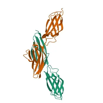
Streptavidin is a 60 kDa bacterial protein with four high‑affinity binding sites for biotin (affinity constant ~10¹⁵ L/mol). Unlike avidin, streptavidin has a neutral isoelectric point and lacks carbohydrate moieties, resulting in significantly lower non‑specific binding in diagnostic assays.
Signal Multiplication via Enzyme Conjugates and Complexes
Once the detection antibody is biotinylated, you introduce a streptavidin conjugate carrying multiple signal‑generating molecules. Common examples are streptavidin‑HRP, streptavidin‑alkaline phosphatase, or streptavidin linked to macromolecular europium chelate complexes.
Because each streptavidin binds to one biotinylated antibody and delivers several enzyme molecules or fluorescent reporters to that spot, the density of signal‑generating enzymes per analyte binding event rises dramatically. A single target molecule can be detected far more easily, directly improving the signal‑to‑noise ratio and lowering the detection limit.
Architectures for Signal Amplification
Labeled Avidin‑Biotin (LAB) System
This is the simplest layout: biotinylated antibody binds the target, then streptavidin‑enzyme conjugate binds directly. It provides straightforward signal enhancement with minimal extra steps.
Bridged Avidin‑Biotin (BRAB) System
Here, unmodified streptavidin acts as a bridge. It first binds the biotinylated antibody, then captures a separately added biotinylated enzyme. Because streptavidin has multiple open sites, it can tether extra reporter molecules, yielding more signal than a direct LAB setup.
Avidin‑Biotin Complex (ABC) System
Biotinylated enzymes and streptavidin are pre‑mixed in precise ratios to form high‑molecular‑weight multimeric complexes that still possess unoccupied biotin‑binding sites. When this complex attaches to a biotinylated antibody, it delivers a massive concentration of enzyme labels per binding event, offering the highest amplification among standard immunoassay formats.
Solving Real‑World Challenges
Overcoming Steric Hindrance in Rapid Assays
In rapid kinetic immunoassays (e.g., 25‑minute protocols), binding often occurs under non‑equilibrium conditions that can limit signal. Steric hindrance around the biotin binding site further impairs efficiency.
The solution combines biotin derivatives with an extended 14‑atom spacer and streptavidin‑polyperoxidase conjugates. The spacer relieves steric crowding and allows high‑affinity binding to proceed quickly. The polyperoxidase polymer then delivers multiple active enzyme molecules per streptavidin, amplifying signal even when incubation times are cut short.
Mitigating Biotin Interference from Patient Samples
High concentrations of free biotin in patient samples compete with biotinylated reagent components for streptavidin binding sites. This can cause false negatives in sandwich assays (reduced capture) or false positives in competitive assays (uncaptured tracer).
To mitigate interference during IVD development, you can:
- Incorporate biotin‑scavenging pre‑incubation steps.
- Add anti‑biotin neutralizing antibodies to assay buffers.
- Switch to biotin‑free conjugation chemistries (direct covalent coupling) where possible.
- Use streptavidin‑coated magnetic beads or solid‑phase pre‑treatment materials to deplete excess biotin from the sample before the assay.
Understanding the Trade‑offs
- Biotin interference is real. Any assay relying on streptavidin‑biotin binding must be validated for its biotin interference threshold, particularly when targeting low‑abundance analytes where small signal losses can matter.
- The highest‑amplification formats (ABC) add complexity. Pre‑forming complexes and maintaining precise reagent ratios demands tighter manufacturing controls but buy you unparalleled sensitivity.
- Streptavidin is preferred over avidin. Although avidin has similar binding strength, its glycosylation and basic pI lead to higher background signals, which can mask the gains you’re trying to achieve for trace targets.
- Not every antibody will tolerate multiple biotin labels. While gently, some monoclonals can lose affinity if critical primary amines are over‑modified; reagent optimization is essential.
Making the Right Choice for Your Sensitivity Goal
Your amplification strategy should match your diagnostic product’s core need. Here’s how to prioritize:
- If your primary focus is maximum signal per binding event: Adopt the ABC system to deliver a pre‑formed polymer of enzyme labels at every target site.
- If your primary focus is reducing assay time while maintaining sensitivity: Combine an extended‑spacer biotin derivative with a streptavidin‑polyperoxidase conjugate to overcome steric hindrance and boost signal under non‑equilibrium conditions.
- If your primary focus is building a flexible IVD platform: Use streptavidin‑coated microplates or magnetic beads as a universal capture phase—any biotinylated antibody can be plugged in, eliminating unique solid‑phase chemistry for each new analyte.
- If your primary focus is avoiding biotin interference: Evaluate direct covalent conjugation of your detection antibodies or implement a validated biotin‑scavenging pre‑treatment step to safeguard assay accuracy.
Leveraging biotin‑streptavidin as an IVD raw material strategy doesn’t just amplify signal—it gives you a modular, scalable architecture that can make the difference between missing a low‑abundance biomarker and detecting it with confidence.
Summary Table:
| Amplification Strategy | Architecture / Mechanism | Primary Advantage | Best Use Case |
|---|---|---|---|
| LAB System | Biotinylated antibody + Streptavidin-reporter conjugate | Simple 2-step setup with minimal protocol complexity | Standard immunoassays needing moderate signal boost |
| BRAB System | Biotinylated antibody + Unlabeled Streptavidin + Biotinylated reporter | Streptavidin bridge tethers additional enzyme labels | Assays requiring higher signal than direct LAB |
| ABC System | Biotinylated antibody + Pre-formed Streptavidin-enzyme complexes | Massive concentration of enzyme labels per binding event | Maximum sensitivity for ultra-low abundance targets |
| Extended Spacer + Poly-HRP | 14-atom spacer biotin + Streptavidin-polyperoxidase | Overcomes steric crowding & boosts kinetics under non-equilibrium | Rapid diagnostic assays with shortened incubation times |
Maximize Assay Sensitivity with CamelBio
Struggling to boost signal density or eliminate background interference in your diagnostic assays? CamelBio provides diagnostic manufacturers, labs, and research institutes with one-stop access to IVD raw materials, technical services, and consulting—covering every stage from concept to clinic.
From high-performance streptavidin reagents to custom conjugation and assay optimization, our experts are ready to accelerate your IVD development.
Partner with CamelBio today to unlock superior assay performance!
Related Products
- APC Streptavidin
- Streptavidin Monoclonal Antibody for Flow Cytometry - SA V1, SA V2
- Streptavidin
- Biotin Rabbit Polyclonal Antibody
- APC Streptavidin Monoclonal Antibody for Flow Cytometry - APC Streptavidin
People Also Ask
- What are key chemical steps to prepare HRP-streptavidin conjugates via periodate oxidation? Optimize IVD performance
- What heterobifunctional strategy conjugates streptavidin with HRP/AP lacking native SH? Master Sulfo-SMCC & SATA.
- How does biotin-(strept)avidin coupling enhance detection sensitivity? Maximize Immunoassay Signal Density
- How are carrier proteins and hapten conjugates utilized to develop light-scattering inhibition immunoassays for small-molecule analytes?
- What role do labeled probes and conjugate enzymes play in nucleic acid hybridization assays? Signal & Specificity