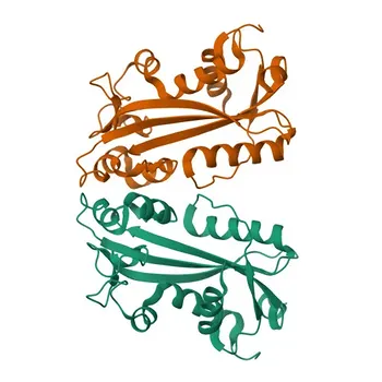
The role of labeled probes and conjugate enzymes is to transform an invisible, molecular binding event into a detectable, quantifiable signal. A labeled probe acts as a highly specific "homing beacon" that finds and binds only its complementary target nucleic acid sequence. The conjugate enzyme, often linked to that probe, then acts as a powerful "amplifier," catalyzing a reaction that produces many millions of localized signal molecules—light, color, or fluorescence—enabling even single-copy gene detection in a background of complex biological material.
While the probe provides the recognition, the conjugate enzyme provides the sensitivity. Together, they solve the core challenge of nucleic acid assays: detecting a needle (the target) in a haystack (the sample) with the certainty required for clinical decisions.
The Core Components of Detection
Understanding the distinct roles of the probe and the enzyme reveals why this two-part system is the foundation of modern molecular diagnostics, from traditional Southern blots to high-density microarrays.
Labeled Probes: The Specificity Engine
The probe's job is molecular identity. It is a short, single-stranded piece of nucleic acid designed to be the exact complement of your target gene or sequence.
Because Watson-Crick base pairing is so precise, a well-designed probe will only hybridize to its perfect match. This specificity is what allows you to ask a single question in a human genome containing billions of base pairs.
The probe also carries the "label"—such as a fluorophore (for direct detection) or a biotin molecule (for indirect detection). This label is the first link in the signal chain, the antenna that will later draw in the enzyme.
Enzyme Conjugates: The Signal Amplifier
The conjugate enzyme’s job is signal multiplication. A single probe-target binding event is not loud enough to detect on its own. The enzyme solves this.
In a common biotin-streptavidin system, the probe’s biotin label recruits a streptavidin-enzyme conjugate (e.g., streptavidin-HRP or streptavidin-AP). Streptavidin binds biotin with near-covalent strength, creating a rock-solid bridge.
Once anchored, each enzyme molecule can catalyze the turnover of thousands of chemiluminescent, colorimetric, or fluorescent substrates per second. This catalytic amplification turns a single, silent hybridization event into a pile of photons that a camera or scanner can easily capture.
Why This System Is Indispensable for Clinical Assays
The biological samples used in Southern blotting or microarray analysis are notoriously difficult to work with. The probe-enzyme system directly addresses these real-world challenges.
Overcoming Sensitivity Limitations
A standard nucleic acid stain like ethidium bromide may only detect DNA in the nanogram range. A target-specific labeled probe can deliver up to 125-fold higher sensitivity, detecting as little as 0.1 picograms of DNA.
This extreme sensitivity is not a luxury. It is the difference between missing a low-abundance pathogen early in an infection and identifying it in time to act. It enables the detection of rare mRNA transcripts in a microarray, revealing subtle gene expression patterns driving a disease.
Conquering Background Noise and Interference
Clinical samples contain autofluorescent debris, endogenous enzymes, and molecules like endogenous biotin that can create false-positive signals.
The enzyme-substrate system provides a superior signal-to-noise ratio. The generated signal is so intense and localized that it stands out sharply against a dark background. Furthermore, diagnostic platforms can use tailored blocking steps and alternative linker systems (biotin-free) to eliminate interference, ensuring the signal you see comes only from your specific target.
Understanding the Trade-offs
No detection technology is without limitations. Recognizing these helps you troubleshoot and design better experiments.
- Non-Specific Binding: The streptavidin-biotin system is powerful, but streptavidin can stick non-specifically to certain sample components, contributing to background. Proper blocking is essential.
- Endogenous Biotin Interference: Tissues like liver or kidney are rich in endogenous biotin, which will bind the conjugate even without your probe. Moving to a digoxigenin/anti-digoxigenin system or using direct fluorophore-labeled probes can circumvent this entirely.
- Enzyme Sensitivity: HRP is susceptible to inactivation by azide preservatives or extreme pH. Alkaline phosphatase requires a specific buffer environment. Choosing the wrong enzyme for your substrate compatibility can extinguish your signal.
Making the Right Choice for Your Goal
Your application dictates how you should pair the probe and enzyme. Align the technology with the problem you are solving.
- If your primary focus is maximum sensitivity for low-copy targets: Prioritize an indirect detection system using a biotinylated probe and a streptavidin-HRP conjugate with an ultra-sensitive chemiluminescent substrate. The amplification factor is unmatched.
- If your primary focus is quantitative microarray analysis: Use direct fluorophore-labeled probes or a single-step indirect system (e.g., with a fluorescent streptavidin conjugate). This provides a linear signal directly proportional to target abundance, avoiding the saturation risks of enzymatic amplification.
- If your primary focus is diagnosing a pathogen in problematic tissue (e.g., biopsies): Avoid the streptavidin-biotin lock entirely. Use a probe labeled directly with alkaline phosphatase or a hapten like digoxigenin to eliminate background from endogenous biotin.
The probe asks the right question, and the enzyme shouts the answer. Mastering their interplay turns a theoretical binding event into a diagnosis you can trust.
Summary Table:
| Component | Primary Role | Key Mechanism | Best Used For |
|---|---|---|---|
| Labeled Probe | Specific Target Recognition | Base pairing (Watson-Crick) with complementary target DNA/RNA | Sequence identification, single-base specificity |
| Conjugate Enzyme | Signal Amplification | Catalyzes substrate turnover to emit light, color, or fluorescence | Detecting low-abundance/single-copy targets |
| Direct Probe System | Rapid, Quantitative Signal | Direct signal emission from attached fluorophore | High-density microarrays, linear quantitation |
| Indirect Enzyme System | Maximum Sensitivity | Streptavidin/biotin or DIG enzyme recruitment (HRP/AP) | Trace detection in complex clinical samples |
Accelerate Your Molecular Diagnostic Assays with CamelBio
Whether you are developing high-sensitivity hybridization protocols or optimizing microarrays, CamelBio provides diagnostic manufacturers, labs, and research institutes with one-stop access to IVD raw materials, technical services, and consulting—covering every stage from concept to clinic.
Ready to elevate your assay performance? Contact our expert technical team today to explore high-quality enzymes, specialized linkers, and custom diagnostic solutions.
Related Products
- APC Streptavidin
- Rabbit anti-FITC/5-FAM/6-FAM mAb - FITC
- Monoclonal Mouse IgG1 Isotype Control Antibody (Conjugate) for Flow Cytometry - Mouse IgG 1 isotype control
- Rabbit anti-FITC/5-FAM/6-FAM Monoclonal Antibody - FITC
- Anti-ITPA Polyclonal Antibody for WB, ELISA - Q9BY32
People Also Ask
- How can diagnostic developers utilize biotin-streptavidin reagents to amplify signals for low-abundance targets?
- What are key chemical steps to prepare HRP-streptavidin conjugates via periodate oxidation? Optimize IVD performance
- What heterobifunctional strategy conjugates streptavidin with HRP/AP lacking native SH? Master Sulfo-SMCC & SATA.
- How does biotin-(strept)avidin coupling enhance detection sensitivity? Maximize Immunoassay Signal Density
- How are carrier proteins and hapten conjugates utilized to develop light-scattering inhibition immunoassays for small-molecule analytes?