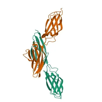
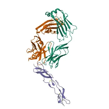
Biotin-(strept)avidin creates a high-density signaling scaffold. The biotin-(strept)avidin coupling system elevates immunoassay sensitivity by enabling multiple indicator labels—enzymes or fluorophores—to accumulate at a single antigen-antibody binding event. In an indirect assay format, a biotinylated secondary antibody recognizes the primary antibody, then a (strept)avidin conjugate carrying the detection label binds with femtomolar affinity. Because (strept)avidin possesses four biotin-binding sites and antibodies can be tagged with several biotin molecules, the system recruits far more signaling units than a directly labeled antibody could carry, dramatically amplifying chromogenic, chemiluminescent, or fluorescent output and lowering the limit of detection.
The biotin-(strept)avidin system’s power lies in its multivalency: one antibody can recruit many enzyme- or fluorophore-linked (strept)avidin molecules, converting a single molecular recognition event into a cascade of detectable signal. Mastering biotinylation ratios and conjugate purity is essential to harness this amplification without introducing steric interference or background.
The Molecular Mechanism of Signal Amplification
The Affinity and Stoichiometry That Drive Sensitivity
The interaction between biotin and (strept)avidin is one of the strongest non-covalent bonds in nature (K_D ≈ 10⁻¹⁵ mol/L). This near-irreversible binding ensures that once the biotinylated antibody is attached to its target, the subsequent (strept)avidin-conjugate step is extraordinarily efficient.
Streptavidin has four biotin-binding sites, so each bound protein can still capture additional biotinylated molecules. Meanwhile, a single antibody can be conjugated with multiple biotins (typically 4–8 per IgG) without losing immunoreactivity. The combination creates a web where many (strept)avidin-label complexes dock onto one antibody-antigen site.
How the Indirect Assay Stack Builds a Label-Rich Complex
In a typical indirect immunoassay, the target antigen is immobilized on a solid phase. The primary antibody binds, and then a biotinylated secondary antibody attaches to the primary’s Fc region. A preformed (strept)avidin-indicator complex—such as streptavidin-HRP (horseradish peroxidase) or streptavidin-alkaline phosphatase—is introduced.
The resulting scaffold multiplies the number of enzyme or fluorophore labels per binding event. When the enzyme substrate is added, each enzyme molecule catalyzes the conversion of many substrate molecules, creating a powerful signal that correlates with the target concentration. This layered architecture is what pushes sensitivity into the picogram-per-milliliter range.
Raw Material Development: Critical Quality Attributes
Precise Biotinylation Ratios Are Everything
The biotinylation step of the secondary antibody must be tightly controlled. Too few biotins limit amplification because fewer (strept)avidin conjugates can bind. Too many biotins can create steric hindrance, masking the antibody’s antigen-binding site or causing it to adopt a conformation that reduces affinity.
For maximum sensitivity, raw material developers typically aim for 4–6 biotins per IgG molecule, achieved through careful reagent ratios and reaction monitoring. Validating the degree of biotinylation via HABA assay or mass spectrometry ensures batch-to-batch consistency.
High-Purity (Strept)avidin–Enzyme Conjugates
The (strept)avidin-enzyme conjugate itself must be of high purity to avoid noisy backgrounds. Free enzyme or free streptavidin in the preparation can compete for binding or generate nonspecific signal. Using pre-formed complexes where streptavidin is incubated with biotinylated enzyme under optimized molar ratios yields large, active polymers with minimal free monomer.
This is the foundation of the Avidin-Biotin Complex (ABC) method, where the complex can contain dozens of enzyme molecules per streptavidin core. High-quality conjugates ensure that every added label actively participates in signal generation.
Comparing Amplification Configurations
Labeled Avidin-Biotin (LAB): Direct but Limited
In the LAB method, a streptavidin–enzyme conjugate is added after the biotinylated antibody. Each streptavidin carries a few enzyme molecules directly attached. While this still amplifies beyond a directly labeled primary antibody, the enzyme density per binding event is moderate.
Bridged Avidin-Biotin (BRAB): Adding Layers
BRAB introduces a native (unlabeled) avidin or streptavidin as a bridge between the biotinylated antibody and a separate biotinylated enzyme. This creates an extra layer, but the stoichiometry is less controlled and the signal gain may not match ABC.
Avidin-Biotin Complex (ABC): The High-Sensitivity Powerhouse
The ABC system pre-assembles a polymeric matrix by mixing (strept)avidin with biotinylated enzyme in specific proportions. Because streptavidin has four biotin-binding sites, it forms a three-dimensional network loaded with active enzyme molecules. When this pre-formed complex is introduced to the biotinylated antibody, a single binding event delivers a massive, multi-enzyme cluster, producing signal amplification that can exceed a thousand-fold per biotin label. This makes ABC reagents ideal for ultra-sensitive ELISA and immunohistochemistry applications.
Understanding the Trade-offs and Common Pitfalls
When Over-Biotinylation Becomes a Problem
Steric hindrance can prevent the biotinylated antibody from effectively binding its target or can block the (strept)avidin conjugate’s access to the biotin sites. This reduces effective signal and may paradoxically lower sensitivity. Careful biotinylation optimization—balancing tag density with retained biological activity—is non-negotiable.
Navigating Endogenous Biotin Interference
Many biological samples (especially liver, kidney, and serum) contain endogenous biotin or biotin-binding proteins. Without proper blocking steps, the (strept)avidin conjugate may bind to these native biotin groups, increasing background and false positives. Using streptavidin (which has lower nonspecific binding than avidin) and pre-blocking with free avidin/biotin can mitigate this.
Conjugate Aggregation and Lot Consistency
The ABC complex is sensitive to molar ratios. Slight deviations can lead to large aggregates that precipitate or bind nonspecifically. For kit manufacturing, each lot of streptavidin–enzyme conjugate must be rigorously validated for size distribution and activity. Even small lot-to-lot variations can shift assay sensitivity and reproducibility.
Making the Right Choice for Your Assay
Tailoring the biotin-(strept)avidin approach to your specific diagnostic goals ensures you maximize sensitivity while maintaining practicality.
- If your primary focus is maximum sensitivity for low-abundance targets: Use the Avidin-Biotin Complex (ABC) configuration with high-purity streptavidin and a biotinylated secondary antibody optimized for ~4–6 biotins per IgG. This combination generates the highest signal density per analyte molecule.
- If you prioritize rapid development and simplicity with good sensitivity: The Labeled Avidin-Biotin (LAB) method offers a straightforward workflow while still delivering more signal than a directly labeled primary antibody.
- If you need to minimize background in complex matrices: Choose streptavidin over avidin, block endogenous biotin with a biotin-blocking kit, and fine-tune biotinylation to avoid excess free biotin or over-modified antibodies that increase nonspecific sticking.
- If reproducibility and scalability are key: Invest in rigorously controlled raw materials—streptavidin–enzyme conjugates with defined molar substitution ratios and validated antibody biotinylation levels. This ensures lot-to-lot consistency, which is critical for IVD kit performance.
By engineering the biotin-(strept)avidin molecular scaffold with precision, you transform a single, specific binding event into a robust, quantifiable signal—pushing your immunoassay’s detection limits to new heights.
Summary Table:
| Configuration | Mechanism | Amplification Level | Ideal Use Case |
|---|---|---|---|
| LAB (Labeled Avidin-Biotin) | Direct binding of streptavidin-enzyme conjugate to biotinylated antibody | Moderate | Rapid development and simplified assay workflows |
| BRAB (Bridged Avidin-Biotin) | Native streptavidin bridges biotinylated antibody and biotinylated enzyme | Moderate to High | Custom multi-layered assay architectures |
| ABC (Avidin-Biotin Complex) | Pre-formed 3D matrix of streptavidin and biotinylated enzymes | Maximum (>1000-fold) | Ultra-sensitive ELISA & IHC for low-abundance analytes |
Ready to optimize your immunoassay sensitivity and secure reliable IVD raw materials? CamelBio provides diagnostic manufacturers, labs, and research institutes with one-stop access to high-purity IVD raw materials, technical services, and consulting—covering every stage from concept to clinic. Whether you are fine-tuning biotinylation ratios or scaling up conjugate manufacturing, contact us today to collaborate with our experts!
Related Products
- Streptavidin
- APC Streptavidin
- APC Streptavidin Monoclonal Antibody for Flow Cytometry - APC Streptavidin
- Anti-CD31/PECAM1 Monoclonal Antibody (Biotin) for FC - Q08481
- PE Streptavidin Monoclonal Antibody (Rabbit) - SA V1, SA V2
People Also Ask
- What are key chemical steps to prepare HRP-streptavidin conjugates via periodate oxidation? Optimize IVD performance
- How can diagnostic developers utilize biotin-streptavidin reagents to amplify signals for low-abundance targets?
- What heterobifunctional strategy conjugates streptavidin with HRP/AP lacking native SH? Master Sulfo-SMCC & SATA.