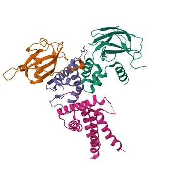
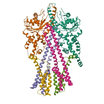
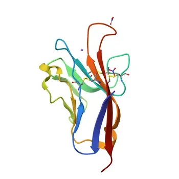
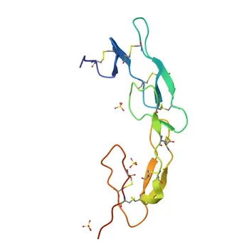
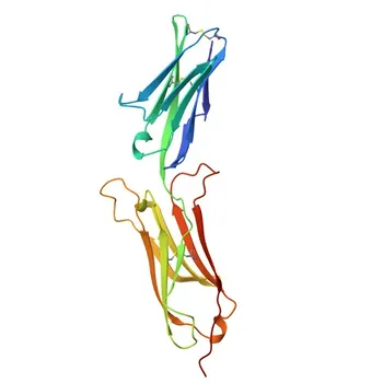
Camelid VHH domains solve the intrinsic stability, production, and targeting limitations of conventional antibodies by design. Unlike mammalian IgGs and their heterodimeric fragments (scFv/Fab), VHH reagents consist of a single, unpaired variable domain that eliminates chain-pairing complexity. Their unique architecture—featuring hydrophilic substitutions at the former light-chain interface—dramatically reduces aggregation, enables high-yield bacterial expression, and delivers exceptional thermal and chemical resilience. This produces IVD raw materials with extended shelf‑life, robust performance, and the ability to bind recessed or cryptic epitopes that remain invisible to bulkier antibody formats.
The shift to VHH reagents isn’t a trend—it’s a direct response to the diagnostic field’s need for recombinant binders that combine thermodynamic stability, production simplicity, and epitope access that IgGs and scFvs simply cannot match.
The Hidden Demands of Modern IVD Development
Diagnostic assay developers face a tension that conventional antibody tools rarely resolve: reagents must be exquisitely stable yet easy to manufacture, sensitive yet rugged enough for harsh matrices. A successful IVD raw material must perform consistently across thousands of tests, tolerate shipping and storage without cold-chain fragility, and often survive regeneration cycles in automated instruments. Any weakness in these areas destabilizes lot‑to‑lot consistency and erodes clinical reliability.
Why Conventional IgGs and Fragments Fall Short
Full‑length mammalian IgGs are large (~150 kDa), require complex glycosylation, and depend on correct pairing of two distinct chains. Their production in mammalian cells is expensive and time‑consuming, and their stability is often conditioned on a narrow physicochemical window.
scFv fragments were designed to address some of those hurdles, but they introduce new problems. The synthetic linker that holds the variable domains together is prone to degradation, leading to aggregation and inclusion‑body formation during recombinant expression. Critically, scFv molecules frequently lose binding function following covalent immobilization or chemical conjugation—a fatal flaw for biosensor‑based or label‑dependent IVD formats.
The VHH Solution: Architecture That Meets the Need
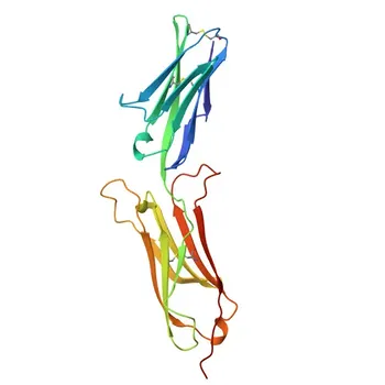
Camelid single‑domain antibodies (also called nanobodies or VHH) bypass these limitations at the molecular level. They are naturally devoid of light chains and the CH1 domain, existing as a single ~14‑15 kDa variable heavy‑chain domain. This stripped‑back design eliminates the need for chain pairing, linkers, or eukaryotic processing, allowing them to function as extraordinarily stable, fully recombinant binding units that can be produced in simple bacterial systems. Every feature that makes a diagnostic reagent hard to scale or hard to trust is architecturally absent from VHH domains.
How VHH Architecture Transforms Reagent Performance
The diagnostic advantage of VHH reagents flows directly from a handful of biophysical traits that conventional formats lack.
Stability That Redefines Shelf‑Life
VHH domains exhibit exceptional thermodynamic stability and a high capacity to refold after thermal stress. They resist denaturation by surfactants, organic solvents, and extreme pH, thanks in part to an additional intra‑chain disulfide bond that locks the CDR1 and CDR3 loops into a robust configuration. In practical terms, this means a VHH‑based assay component can be shipped without cold chain, stored for years with minimal activity loss, and deployed in point‑of‑care devices that endure wide environmental fluctuations.
Aggregation‑Proof and Super‑Soluble
At the former light‑chain interface, VHH sequences carry hallmark hydrophobic‑to‑hydrophilic amino acid substitutions. These substitutions shield the hydrophobic core that would otherwise be exposed in a single‑domain binder, dramatically reducing aggregation and enabling high‑yield soluble expression in E. coli and yeast. Unlike scFv, VHH reagents require no synthetic linker, eliminating the primary source of linker‑driven aggregation and inclusion‑body formation. The result is a raw material that can be purified at scale with minimal loss and formulated into homogeneous, stable liquid reagents.
Accessing Cryptic and Cavity Epitopes for Superior Specificity
The extended CDR3 loop of a VHH—often 16‑18 amino acids long—can form a finger‑like projection that penetrates narrow protein clefts, enzymatic active sites, and recessed epitopes that are physically inaccessible to the larger, flatter paratopes of conventional IgGs or scFvs. This ability to target hidden or cavity‑bound epitopes unlocks diagnostic specificity that bulky antibody formats cannot achieve, whether the goal is detecting a conformational variant of a biomarker or capturing a small molecule deep within a binding pocket.
Ruggedness from Bench to Field
Where scFv molecules often lose function upon surface immobilization or chemical labeling, VHH reagents retain full antigen‑binding activity after covalent attachment to biosensor surfaces or conjugation with detection labels. This ruggedness means a single VHH clone can serve both as a capture and a detection element, simplifying assay architecture. Moreover, immunoaffinity columns and sensor surfaces functionalized with VHHs can withstand thousands of harsh regeneration cycles—including exposure to low pH, high salt, or denaturing solvents—without measurable loss of binding capacity. That durability translates directly into lower per‑test costs and less frequent recalibration.
Understanding the Trade‑offs When Adopting VHH Reagents
No technology is a universal panacea, and VHH reagents come with design considerations that diagnostic developers should weigh from the start.
- Monovalency demands intentional engineering. Native VHHs are monovalent. While this can be an advantage for certain detection modes, sandwich assays that rely on multivalent binding or Fc‑mediated orientation will require intentional multimerization or addition of spacer domains. However, because VHHs are so stable, tandem fusions and oriented immobilization are straightforward to implement compared to scFv or IgG.
- Immobilization orientation still matters. VHHs tolerate a wide range of immobilization chemistries, but random amine coupling can occasionally mask the paratope if critical lysines are positioned near the CDR loops. Site‑specific biotinylation or click‑chemistry strategies are recommended to maximize functional density on sensor surfaces, but the inherent refolding ability of VHHs makes them far more forgiving than scFv.
- Epitope recognition is biased toward cavities. The extended CDR3 loop excels at engaging concave surfaces, but it may be less optimal for flat, featureless epitopes where a conventional Fab or IgG might provide a larger contact area. This is rarely a limitation for high‑specificity IVD applications, but it deserves consideration during lead‑screening strategies.
These are manageable design choices, not fundamental limitations. What VHHs give in return—unmatched stability, production economy, and cryptic‑epitope access—far outweighs the adjustment effort for most IVD programs.
Making the Right Choice for Your IVD Assay
Use the table below—or rather, the following goal‑oriented recommendations—to decide whether VHH reagents align with your diagnostic development strategy.
- If your primary focus is long‑term shelf‑life and field‑ready stability: VHH reagents are the superior choice. Their thermodynamic robustness and resistance to environmental stress eliminate cold‑chain dependency and reduce lot‑to‑lot performance drift.
- If your target biomarker presents a buried, occluded, or enzymatic active‑site epitope: VHH’s extended CDR3 loop can reach epitopes that IgGs and scFvs cannot, enabling specific detection that other formats simply miss.
- If you need to scale production cost‑effectively with batch‑to‑batch consistency: High‑yield expression in bacterial systems, absent complex glycosylation or linker‑driven aggregation, makes VHH manufacturing predictable and economical.
- If your assay platform relies on harsh regeneration or rigorous surface immobilization: VHH domains retain full activity after chemical conjugation and withstand thousands of regeneration cycles without performance loss, a feat no scFv or full‑length IgG can match.
Camelid VHH reagents are not just a replacement for conventional antibodies—they are an architectural upgrade that solves the deep, often hidden pain points of IVD assay development. By aligning your binder tool with the real‑world stresses of diagnostic use, you build assays that are more reliable, more accessible, and ultimately more valuable to the patient.
Summary Table:
| Feature | Camelid VHH Reagents | Conventional IgG | scFv Fragments |
|---|---|---|---|
| Molecular Weight | ~14–15 kDa | ~150 kDa | ~25 kDa |
| Thermal & Chemical Stability | High (refolds readily after stress) | Moderate (cold-chain dependent) | Low to Moderate (prone to aggregation) |
| Expression System | High-yield E. coli / Yeast | Complex Mammalian Systems | E. coli (often forms inclusion bodies) |
| Synthetic Linker Requirement | None (native single domain) | None | Required (causes linker-driven aggregation) |
| Cryptic/Cavity Epitope Access | Superior (extended CDR3 loop) | Poor (large, flat paratope) | Moderate |
| Regeneration Cycle Resilience | Retains activity after 1000s of cycles | Poor | Poor to Moderate |
Upgrade Your Diagnostic Assays with High-Performance VHH Reagents
Ready to eliminate cold-chain bottlenecks and access hidden biomarker targets in your diagnostic platforms? CamelBio provides diagnostic manufacturers, labs, and research institutes with one-stop access to IVD raw materials, technical services, and consulting—covering every stage from concept to clinic.
Whether you need custom VHH development, high-yield recombinant production, or assay optimization, our expert team is here to power your success. Contact CamelBio Today to start your project!
Related Products
- KO Validated Anti-Von Hippel Lindau/VHL Rabbit Monoclonal Antibody for WB, ELISA - P40337
- Anti-CAMLG Rabbit Polyclonal Antibody for WB, IHC-P, IF/ICC, ELISA - P49069
- HAVCR1 Rabbit pAb - Q96D42
- Anti-Mouse CD270/HVEM Monoclonal Antibody for Flow Cytometry - Q80WM9
- Anti-CaMKII delta (CAMKD) Rabbit Monoclonal Antibody for WB, IF/ICC, ELISA - Q13557