Monoclonal Antibodies
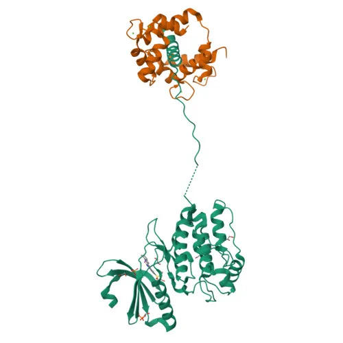
Anti-CaMKII delta (CAMKD) Rabbit Monoclonal Antibody for WB, IF/ICC, ELISA - Q13557
Item Number : CM0004372
Price varies based on specs and customizations
- Application
- WB, IF/ICC, ELISA
- Cross Reactivity
- Human, Mouse
- Protein Weight
- 56 kDa

Shipping:
Contact us to get shipping details Enjoy On-time Dispatch Guarantee.
Why Choose Us
Easy ordering process, quality products, and dedicated support for your business success.
Core Product Specifications and Parameters
| Parameter | Value |
|---|---|
| Product Name | CaMKII delta Rabbit mAb |
| Remarks/Alias | CAMKD; CaMKII delta |
| Species | Human |
| GeneID (Human) | 817 |
| GeneID | 817 |
| Immunogen | Synthetic Peptide: A synthetic peptide corresponding to a sequence within amino acids 1-100 of human CaMKII delta (Q13557) |
| Source | Rabbit |
| Category | Monoclonal Antibodies |
| Application | WB, IF/ICC, ELISA |
| Cross Reactivity | Human, Mouse |
| SWISS | Q13557 |
| Protein Weight | 56 kDa |
| Shipping | Ice bag |
Biological Background: CaMKII delta Function and Localization
- CaMKII delta is a calcium/calmodulin-dependent serine/threonine kinase belonging to the CAMK2 family, encoded by the CAMK2D gene; multiple splice isoforms exist, with isoform Delta 3, 2, 8, 9 expressed in cardiac muscle and Delta 11 in skeletal muscle.
- Serves as a central regulator of cardiac excitation-contraction coupling by targeting ion channels and transporters, including L-type calcium channel CACNB2, sodium channel Nav1.5/SCN5A, and potassium channel Kv4.3/KCND3.
- Mediates SR Ca2+ release by direct phosphorylation of ryanodine receptor RYR2 at Ser-2808, contributing to cardiac decompensation in heart failure.
- Phosphorylates phospholamban (PLN), relieving inhibition of SERCA2A and enhancing SR Ca2+ uptake, which is important for frequency-dependent acceleration of relaxation and contractile function during acidosis. Related references: PMID:16690701
- In the nucleus, phosphorylates HDAC4, promoting its nuclear export and enabling MEF2-driven hypertrophic gene expression. Related references: PMID:17179159
- Subcellularly localized to the sarcolemma and sarcoplasmic reticulum membrane, where it forms macromolecular complexes with its substrates.
- In response to interferon-gamma, catalyzes phosphorylation of STAT1, stimulating the JAK-STAT pathway.
- Post-translationally regulated by acetylation and autophosphorylation; structural studies, ATP-binding, and calmodulin-binding are key functional features.
Experimental Guidance and Technical Tips
- The immunogen is a synthetic peptide spanning amino acids 1–100 of human CaMKII delta; consider using this N-terminal region to differentiate from other CAMK2 family members if specificity is critical.
- For Western blot, the predicted molecular weight is ~56 kDa; validate band patterns in cardiac tissue lysates, and include phosphatase inhibitors to preserve phosphorylation states.
- In immunofluorescence/ICC, anticipate sarcolemmal and sarcoplasmic reticulum staining; appropriate fixation and permeabilization are essential for membrane-bound targets.
- ELISA performance may be improved by using peptide immunogen as a positive control to confirm antibody affinity.
CamelBio: Your One-Stop Sourcing Bridge
CamelBio supports diagnostic and research communities with comprehensive access to IVD raw materials, including validated monoclonal antibodies for cardiac signaling targets like CaMKII delta. We offer one-stop sourcing solutions—from antibody pairs and bulk ancillary reagents to tailored technical consultation—aimed at accelerating your cardiac biomarker and heart failure assay development from concept to clinic.
REQUEST A QUOTE
Our professional team will reply to you within one business day. Please feel free to contact us!