Polyclonal Antibodies
HAVCR1 Rabbit pAb - Q96D42
Item Number : CM0030266
Price varies based on specs and customizations
- Application
- WB, ELISA
- Cross Reactivity
- Human, Mouse, Rat
- Protein Weight
- 39kDa

Shipping:
Contact us to get shipping details Enjoy On-time Dispatch Guarantee.
Why Choose Us
Easy ordering process, quality products, and dedicated support for your business success.
Core Product Specifications and Parameters
| Parameter | Value |
|---|---|
| Product Name | HAVCR1 Rabbit pAb |
| Remarks/Alias | TIM; KIM1; TIM1; CD365; HAVCR; KIM-1; TIM-1; TIMD1; TIMD-1; HAVCR-1 |
| Species | Human |
| GeneID (Human) | 26762 |
| Immunogen | Recombinant fusion protein containing a sequence corresponding to amino acids 21-125 of human HAVCR1 (NP_036338.2) |
| Source | Rabbit |
| Category | Polyclonal Antibodies |
| Application | WB, ELISA |
| Cross Reactivity | Human, Mouse, Rat |
| SWISS | Q96D42 |
| Protein Weight | 39kDa |
| Shipping | Ice bag |
Biological Background: HAVCR1 Function and Localization
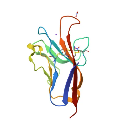
- HAVCR1 encodes a type I transmembrane glycoprotein, also known as Kidney injury molecule 1 (KIM-1), T-cell immunoglobulin and mucin domain-containing protein 1 (TIMD-1), and CD365, containing immunoglobulin and mucin domains.
- The protein functions as a phosphatidylserine receptor that is essential for regulatory B-cell homeostasis, efferocytosis (clearance of apoptotic cells), and phagocytosis of Gram-negative and Gram-positive bacteria. Related references: PMID:18082433 PMID:18414680 PMID:17471468
- In T-cells, HAVCR1 acts as a P-selectin ligand, regulating activated T-cell trafficking and accumulation in the inflamed central nervous system, and controls autoimmune disease susceptibility. Related references: PMID:24703780
- HAVCR1 serves as a cellular receptor for Hepatitis A virus and mediates entry of various enveloped viruses, including Ebolavirus, Marburg virus, Dengue virus, Zika virus, and Chikungunya virus, by binding phosphatidylserine on the virion membrane or interacting with envelope glycoproteins. Related references: PMID:21536871 PMID:26487564 PMID:23084921 PMID:29742433
- Subcellularly, HAVCR1 localizes to the cell membrane, is recruited to the phagocytic cup upon binding apoptotic cells, and is found in phagosomes and cytoplasmic vesicles.
- It is widely expressed, with the highest levels in kidney and testis, and is upregulated on activated CD4+ T-cells during helper T-cell differentiation.
- The protein undergoes glycosylation, disulfide bond formation, isopeptide bond conjugation, and ubiquitination, which may affect its migration in SDS-PAGE.
Experimental Guidance and Technical Tips
- The immunogen comprises amino acids 21-125 of the human HAVCR1 extracellular domain; this antibody may recognize both the full-length membrane protein and shed soluble forms in Western blot and ELISA.
- Predicted molecular weight is 39 kDa, but glycosylation may shift the apparent band to a higher range; consider treating lysates with PNGase F or other deglycosylating enzymes to confirm specificity.
- For Western blot, validate detection in relevant cell lines such as kidney-derived HEK-293 or activated T-cell lines; include positive controls from tissues with high expression (e.g., kidney or testis) to assess cross-reactivity with mouse and rat orthologs.
- In ELISA, this antibody could be used as a capture or detection reagent for quantifying soluble HAVCR1 in serum or cell supernatants; pair with a secondary antibody validated for rabbit IgG.
CamelBio: Your One-Stop Sourcing Bridge
CamelBio streamlines your IVD raw-material sourcing with a comprehensive portfolio spanning infectious disease, kidney injury, and immune function targets. For HAVCR1-related assay development, this polyclonal antibody can be integrated with matched antibody pairs, recombinant antigens, and bulk ancillary reagents available through our global supply network. From research-grade tools to scalable commercial materials, CamelBio supports your project from concept to clinic with reliable, validated solutions.
REQUEST A QUOTE
Our professional team will reply to you within one business day. Please feel free to contact us!