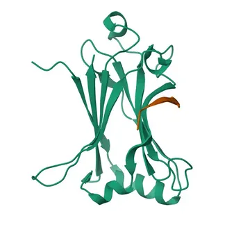
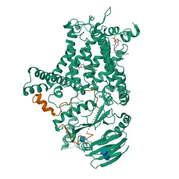
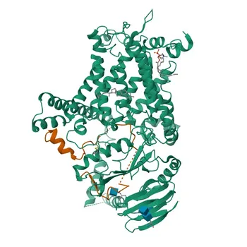
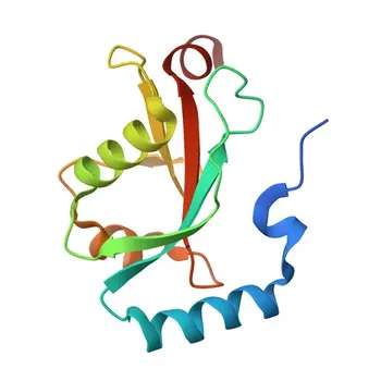
The master architects of bone resorption are the signaling molecules CSF-1 and RANKL, alongside their decoy receptor OPG, which regulate osteoclast formation, while the enzymes TRACP (specifically TRACP5b) and cathepsin K execute the digestion of bone matrix. These molecules can be targeted directly as analytes in immunoassays using high-specificity monoclonal antibodies, or indirectly by measuring the collagen fragments—such as CTX and NTX—that cathepsin K generates, making them invaluable biomarkers for bone metabolism.
The RANKL/OPG balance constitutes the core signaling switch for osteoclast activation, but the most clinically actionable immunoassay targets are the downstream effectors: TRACP5b reflects osteoclast number, and cathepsin K activity is captured by measuring its specific collagen degradation products, CTX and NTX. Successful assay development demands recombinant protein calibrators and antibodies that distinguish the active isoforms from their inactive counterparts.
The Central Signaling Axis: RANKL, OPG, and CSF-1
Understanding the biochemical triggers of osteoclast activation is the first step in selecting a robust analyte. Two key factors orchestrate the entire process from precursor to mature bone-resorbing cell.
CSF-1: The Survival and Recruitment Signal
Colony-stimulating factor 1 (CSF-1), also known as M-CSF, is a non-negotiable growth factor for osteoclast precursor proliferation and survival. It primes the hematopoietic precursors to express the receptor for the next critical signal, RANK.
Without CSF-1, the progenitor pool collapses, making differentiation impossible. In immunoassay development, CSF-1 itself is rarely a direct bone turnover marker because its levels are not as tightly coupled to local bone resorption as the downstream signals. However, its receptor or signaling intermediates remain valuable research targets for understanding osteoclast commitment.
RANKL and OPG: The Differentiation Balance Beam
Receptor activator of nuclear factor-kB ligand (RANKL) binds to its receptor RANK on pre-osteoclasts, triggering the intracellular pathways that drive maturation into multinucleated, active osteoclasts. This single ligand is the non-redundant, essential signal for differentiation and activation.
Osteoprotegerin (OPG) is the natural decoy receptor. It binds RANKL with high affinity, preventing RANKL from engaging RANK. The RANKL/OPG ratio therefore dictates the net bone resorption drive. A high ratio (more RANKL relative to OPG) stimulates osteoclasts; a low ratio protects bone. For an immunoassay developer, this ratio is a crucial diagnostic concept. You can quantify both proteins and calculate the ratio, or measure OPG as a standalone protective signal. The challenge lies in capturing free, bioactive RANKL—much of it is membrane-bound or complexed with OPG in circulation.
The Execution Phase: Enzymes That Degrade Bone
Once an osteoclast attaches to the bone surface, it creates a sealed acidic compartment and deploys its enzymatic arsenal. Two enzymes are the primary candidates for immune-based detection.
Tartrate-Resistant Acid Phosphatase (TRACP5b)
Tartrate-resistant acid phosphatase exists in two isoforms: 5a and 5b. TRACP5b is exclusively osteoclast-derived, released into the circulation during bone resorption. It is not cleared by the liver and has a relatively stable concentration in serum if samples are handled correctly.
Targeting TRACP5b in an immunoassay requires monoclonal antibodies that specifically recognize isoform 5b and not the ubiquitous 5a. This can be achieved by selecting antibodies directed against the unique post-translational modifications or the distinct subunit composition of the active enzyme. A sandwich assay using two such antibodies provides the specificity needed to distinguish osteoclast activity from other tissue sources of acid phosphatase.
Cathepsin K and Its Collagen Fingerprints
Cathepsin K is the predominant cysteine protease secreted into the resorption lacuna. It cleaves the triple-helical type I collagen matrix at specific sites, generating neo-epitope fragments that are excellent targets for an immunoassay.
Rather than measuring the enzyme directly (which can be technically difficult due to inhibitor complexes and low active concentrations), the most successful clinical assays target the products of its activity. The carboxy-terminal telopeptide (CTX) and amino-terminal telopeptide (NTX) are cross-linked peptide fragments released when cathepsin K digests collagen. These fragments are stable, abundant in serum and urine, and highly correlated with bone resorption rates. An immunoassay for CTX, for example, uses a monoclonal antibody specific to the octapeptide sequence EKAHD-β-GGR, which is generated only by cathepsin K cleavage.
Leveraging Collagen Breakdown Products in Immunoassay Design
The supplementary resorption markers—CTX, NTX, DPD, and PYR—all derive from the organic matrix destruction downstream of enzyme activation. They differ in their tissue specificity, sample matrix suitability, and susceptibility to confounding factors.
Direct Enzyme Targets vs. Activity Byproducts
A basic principle of immunoassay development is choosing between measuring the actor or the footprint. For osteoclasts, the enzymes TRACP5b and cathepsin K represent the actors. The telopeptides CTX and NTX represent the footprints.
Direct enzyme measurement (TRACP5b) offers cellular specificity—it tells you how many active osteoclasts are present. Activity byproduct measurement (CTX) integrates the total degradation rate but can be influenced by non-skeletal sources (diet, kidney function). A robust immunoassay panel often includes both types to provide a complete picture of bone metabolism.
Pyridinium Crosslinks: DPD and PYR
Deoxypyridinoline (DPD) and pyridinoline (PYD) are intermolecular crosslinks that stabilize mature collagen fibrils. When bone is resorbed, these crosslinks are released and excreted in urine. While they can be measured by competitive immunoassays using monoclonal antibodies, they have largely been supplanted by serum CTX and NTX assays due to the latter’s superior specificity, ease of specimen handling, and lower biological variability.
For an assay developer, understanding this market trend is important: the investment case for a novel DPD assay is weak unless it offers a significant advantage, like point-of-care urine testing with automated normalization to creatinine.
Understanding the Trade-offs in Assay Development
Every analyte choice comes with a balance of sensitivity, specificity, and practical feasibility. Ignoring these trade-offs leads to assays that fail in clinical validation.
The RANKL Measurement Dilemma
Measuring RANKL seems ideal because it is the central regulator. However, it is a challenging analyte. Most RANKL circulates bound to OPG or is retained on the surface of T-cells and osteoblasts. A total RANKL immunoassay may not reflect the bioactive, free RANKL pool. A specific free-RANKL assay is extremely difficult to standardize because any sample manipulation can shift the binding equilibrium. For routine clinical chemistry, the RANKL/OPG ratio often remains a research tool rather than a frontline diagnostic analyte.
TRACP5b Stability and Sample Integrity
TRACP5b is a heat-labile enzyme that degrades quickly if serum samples are left at room temperature for several hours. Intravenous bisphosphonates can transiently suppress TRACP5b within 24 hours, so sampling timing is critical. An assay kit must include strict instructions for sample collection (e.g., from fasting patients, immediate centrifugation and freezing) and validation against these confounders to gain clinical acceptance.
CTX: Sensitivity at the Cost of Intra-Individual Variability
Serum CTX is exquisitely sensitive to antiresorptive therapy and provides a rapid indication of treatment efficacy. However, it exhibits strong circadian rhythms (highest in the early morning) and is suppressed by food intake. To standardize this marker, the Clinical Laboratory Standards Institute recommends a fasting morning blood draw. An immunoassay kit for CTX must explicitly communicate these pre-analytical requirements, and the assay’s precision profile must be tight enough to detect the 40-60% drop in CTX typical of successful therapy.
Making the Right Choice for Your Assay Development Goal
The selection of biomarker targets depends entirely on the clinical question your immunoassay intends to answer. The following recommendations tie each goal to the most logical analyte choice based on the underlying bone biology.
- If your primary focus is monitoring response to antiresorptive drugs (e.g., bisphosphonates, denosumab): Use a serum CTX immunoassay, as it provides the most sensitive and clinically validated dynamic range for detecting a rapid decrease in bone resorption.
- If your primary focus is assessing renal osteodystrophy or correcting for non-skeletal degradation: Include a TRACP5b assay, because this enzyme is not cleared by the kidneys and specifically reflects the number of active osteoclasts, independent of the collagen source.
- If your primary focus is establishing the central regulatory drive for bone resorption in an untreated population: Develop a panel measuring both RANKL and OPG to calculate the free RANKL index or ratio, confirming that your assay differentiates free active RANKL from bound forms.
- If your primary focus is a low-cost, rapid point-of-care test for overall bone resorption rate: Target NTX in urine using a competitive immunoassay format, but carefully validate against hydration status by normalizing to creatinine.
- If your primary focus is building a comprehensive bone metabolism panel: Pair a resorption marker (such as CTX or TRACP5b) with a formation marker (such as PINP or bone ALP) to capture both sides of the remodeling cycle and provide a full picture of bone turnover balance.
A definitive bone metabolism immunoassay is never about measuring a single molecule in isolation; it is about capturing a clear, stable signal from a highly specific biochemical pathway and delivering a result that aligns precisely with the underlying osteoclast biology.
Summary Table:
| Target Analyte | Category | Biological Function | Immunoassay Strategy & Application |
|---|---|---|---|
| RANKL / OPG | Signaling Axis | Regulates osteoclast differentiation and activation drive | Measure ratio or OPG; requires mAbs distinguishing free active RANKL from bound forms |
| TRACP5b | Resorption Enzyme | Secreted exclusively by osteoclasts during active resorption | Directly reflects osteoclast cell count; requires isoform 5b-specific monoclonal antibody pairs |
| Cathepsin K (CTX / NTX) | Protease / Matrix Byproducts | Cleaves type I collagen matrix during bone digestion | CTX/NTX serve as highly sensitive, clinically validated serum/urine markers for resorption rate |
| CSF-1 (M-CSF) | Growth Factor | Essential for osteoclast precursor proliferation & survival | Primary research target for osteoclast commitment; less sensitive to acute turnover changes |
Accelerate Your Bone Metabolism Immunoassay Development with CamelBio
Developing high-specificity assays for challenging bone turnover targets—such as TRACP5b, CTX, or free RANKL—demands superior antibody specificity and reliable recombinant calibrators.
CamelBio provides diagnostic manufacturers, labs, and research institutes with one-stop access to premium IVD raw materials, technical services, and consulting—covering every stage from concept to clinic. Whether you are scaling production or developing a novel diagnostic panel, we provide the quality reagents and expert support you need to succeed.
👉 Contact CamelBio Today to request raw material samples or consult with our technical team!
Related Products
- Anti-TNFRSF11A Rabbit Polyclonal Antibody for WB, IHC-P, IF/ICC, ELISA - Q9Y6Q6
- Osteocalcin (BGLAP) Rabbit mAb for WB, IF/ICC, ELISA - P02818
- Human Activated T Cell Panel - Rabbit Monoclonal Antibody
- Anti-Osteocalcin (BGLAP) Rabbit Polyclonal Antibody for WB, IF/ICC, ELISA - P02818
- Anti-LC3A/LC3B Rabbit PolymAb® for WB, IHC-P, IF/ICC, ELISA - Q9GZQ8 / Q9H492