Monoclonal Antibodies
ZAP70 Rabbit Monoclonal Antibody for WB, IF-P, ELISA - P43403
Item Number : CM0005184
Price varies based on specs and customizations
- Application
- WB, IF-P, ELISA
- Cross Reactivity
- Human
- Protein Weight
- 70kDa

Shipping:
Contact us to get shipping details Enjoy On-time Dispatch Guarantee.
Why Choose Us
Easy ordering process, quality products, and dedicated support for your business success.
Core Product Specifications and Parameters
| Parameter | Value |
|---|---|
| Product Name | ZAP70 Rabbit mAb |
| Remarks/Alias | SRK; STD; TZK; STCD; IMD48; ADMIO2; ZAP-70; ZAP70 |
| Species | Human |
| GeneID (Human) | 7535 |
| GeneID | 7535 |
| Immunogen | Recombinant fusion protein containing a sequence corresponding to amino acids 550-619 of human ZAP70 (NP_001070.2). |
| Source | Rabbit |
| Category | Monoclonal Antibodies |
| Application | WB, IF-P, ELISA |
| Cross Reactivity | Human |
| SWISS | P43403 |
| Protein Weight | 70kDa |
| Shipping | Ice bag |
Biological Background: ZAP70 Function and Localization
- Tyrosine-protein kinase ZAP-70 (70 kDa zeta-chain associated protein, Syk-related tyrosine kinase) is encoded by the ZAP70 gene and exists as alternatively spliced isoforms.
- Functions as an essential signaling kinase in the adaptive immune response: regulates T-cell activation, proliferation, differentiation, motility, adhesion, and cytokine expression; also contributes to thymocyte development and primary B-cell activation.
- Upon T-cell receptor (TCR) stimulation, ZAP70 is recruited to phosphorylated ITAM motifs of CD3E and CD3Z at the plasma membrane, where it is activated by LCK-mediated phosphorylation. Activated ZAP70 phosphorylates adaptor proteins LAT and LCP2, initiating downstream signaling cascades. Related references: PMID:7509083
- Mediates TCR complex internalization and degradation by interacting with the E3 ubiquitin ligase CBL and adaptors SLA/SLA2, modulating surface TCR expression and signaling duration.
- Subcellular localization: cytoplasm and cell membrane; translocates to the plasma membrane upon TCR engagement.
- Tissue specificity: expressed in T-cells, natural killer cells, early thymocytes, and pro/pre B-cells.
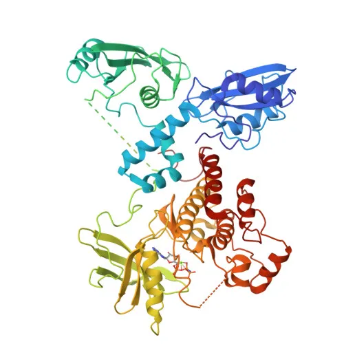
- Post-translational modifications: phosphorylation, acetylation, and ubiquitination regulate its activity and stability. Contains SH2 domains and a kinase domain. Keywords: 3D-structure, phosphoprotein, Ubl conjugation, isopeptide bond, disease variant, SCID.
Experimental Guidance and Technical Tips
- The immunogen is a recombinant fusion protein representing amino acids 550-619 within the C-terminal kinase domain of human ZAP70. For Western blot, expect a specific band around 70 kDa in human T-cell lines (e.g., Jurkat) or peripheral blood mononuclear cells (PBMCs).
- For immunofluorescence (IF-P), permeabilization is required to detect intracellular and membrane-associated ZAP70; consider using T-cell activators to enhance signal.
- In ELISA applications, validate antibody performance with recombinant ZAP70 protein or cell lysates from relevant T-cell samples; titrate optimally.
- Reported cross-reactivity is for human. If testing other species, verify sequence conservation of the immunogen region, particularly in the kinase domain.
CamelBio: Your One-Stop Sourcing Bridge
CamelBio provides comprehensive IVD raw material sourcing for diagnostic developers and research institutes, covering immune profiling, lymphoma diagnostics, and T-cell signaling research. For ZAP70-targeted assays, we offer validated antibody pairs, optimized monoclonal and polyclonal antibodies, bulk ancillary reagents, and rare-target raw material procurement. Our one-stop supply chain and technical services streamline your development from concept to clinic.
REQUEST A QUOTE
Our professional team will reply to you within one business day. Please feel free to contact us!