Polyclonal Antibodies
YKL-40/CHI3L1 Rabbit pAb - P36222
Item Number : CM0019026
Price varies based on specs and customizations
- Application
- WB, IF/ICC, IHC-P, ELISA
- Cross Reactivity
- Human, Mouse, Rat
- Protein Weight
- 43 kDa

Shipping:
Contact us to get shipping details Enjoy On-time Dispatch Guarantee.
Why Choose Us
Easy ordering process, quality products, and dedicated support for your business success.
Core Product Specifications and Parameters
| Parameter | Value |
|---|---|
| Product Name | YKL-40/CHI3L1 Rabbit pAb |
| Remarks/Alias | GP39; ASRT7; GP-39; YK-40; YKL40; CGP-39; YKL-40; YYL-40; HC-gp39; HCGP-3P; hCGP-39; YKL-40/CHI3L1 |
| Species | Human |
| GeneID (human) | 1116 |
| GeneID | 1116 |
| Immunogen | Recombinant fusion protein containing a sequence corresponding to amino acids 22-220 of human YKL-40/CHI3L1 (NP_001267.2) |
| Source | Rabbit |
| Category | Polyclonal Antibodies |
| Application | WB, IF/ICC, IHC-P, ELISA |
| Cross Reactivity | Human, Mouse, Rat |
| SWISS | P36222 |
| Protein Weight | 43 kDa |
| Shipping | Ice bag |
Biological Background: YKL-40/CHI3L1 Function and Localization
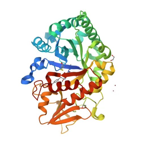
- CHI3L1 (Chitinase-3-like protein 1), also known as YKL-40, GP-39, CGP-39, or HC-gp39, is a secreted glycoprotein belonging to the glycosyl hydrolase 18 family but lacks chitinase catalytic activity.
- It functions as a carbohydrate-binding lectin with a preference for chitin and plays a key role in tissue remodeling and cellular adaptation to environmental changes, particularly in inflammatory conditions.
- CHI3L1 is involved in T-helper cell type 2 (Th2) inflammatory responses and IL-13-induced inflammation, regulating allergen sensitization, inflammatory cell apoptosis, dendritic cell accumulation, and M2 macrophage differentiation.
- In the lung, CHI3L1 regulates antibacterial responses by contributing to macrophage bacterial killing, controlling bacterial dissemination, and augmenting host tolerance. It also mediates hyperoxia-induced injury, inflammation, and epithelial apoptosis.
- Subcellularly, CHI3L1 is primarily secreted into the extracellular space but can also be detected in the cytoplasm, perinuclear region, and endoplasmic reticulum.
- Tissue expression is prominent in activated macrophages, articular chondrocytes, synovial cells, and liver. Expression is very low or undetectable in non-inflammatory colon, muscle, lung, pancreas, mononuclear cells, and fibroblasts.
- The protein undergoes glycosylation and contains a signal peptide and disulfide bonds, which may influence its apparent molecular weight on SDS-PAGE (~43 kDa, higher than the predicted 42.6 kDa).
Experimental Guidance and Technical Tips
- The immunogen spans amino acids 22–220, covering the mature secreted form; the antibody is expected to detect endogenous CHI3L1 in human, mouse, and rat samples.
- For western blotting, a band around 43 kDa is typically observed. Glycosylation and other modifications may cause slight shifts; consider validating with deglycosylation enzymes if precise mass confirmation is required.
- In immunohistochemistry (IHC-P), antigen retrieval may be necessary. Positive controls include inflamed synovial tissue, liver, or activated macrophage-rich samples.
- For IF/ICC, cellular localization (secreted/cytoplasmic) should be confirmed using appropriate fixation and permeabilization protocols. Co-staining with organelle markers can help verify perinuclear or ER localization.
- ELISA applications can be used to quantify CHI3L1 in biological fluids (serum, plasma, culture supernatants); consider pairing with a matched detection antibody if developing a sandwich ELISA.
- Always validate the antibody in your specific sample system and assay conditions, as performance may vary with post-translational modifications and expression levels.
CamelBio: Your One-Stop Sourcing Bridge
CamelBio is a dedicated IVD raw-material partner offering comprehensive access to validated antibody pairs, recombinant proteins, and ancillary reagents for CHI3L1 research and diagnostic development. From monoclonal/polyclonal antibodies to bulk custom formulations, we streamline procurement for inflammation, tissue remodeling, and cancer biomarker studies. For rare targets or scale-up needs in clinical assay manufacturing, CamelBio provides end-to-end sourcing and technical support to bridge the gap from concept to clinic.
REQUEST A QUOTE
Our professional team will reply to you within one business day. Please feel free to contact us!