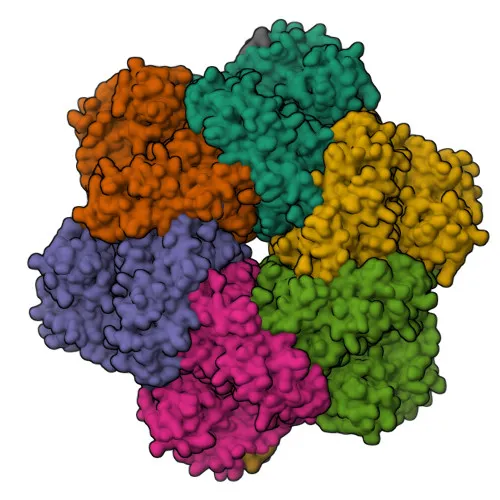
Monoclonal Antibodies
UFD1L Rabbit mAb - Q92890
Item Number : CM0003826
Price varies based on specs and customizations
- Application
- WB, ELISA
- Cross Reactivity
- Human, Rat
- Protein Weight
- 35kDa

Shipping:
Contact us to get shipping details Enjoy On-time Dispatch Guarantee.
Why Choose Us
Easy ordering process, quality products, and dedicated support for your business success.
Core Product Specifications and Parameters
| Parameter | Value |
|---|---|
| Product Name | UFD1L Rabbit mAb |
| Remarks/Alias | UFD1L |
| Species | Human |
| Gene ID (Human) | 7353 |
| Gene ID | 7353 |
| Immunogen | Synthetic peptide |
| Source | Rabbit |
| Category | Monoclonal Antibodies |
| Application | WB, ELISA |
| Cross Reactivity | Human, Rat |
| SWISS | Q92890 |
| Protein Weight | 35kDa |
| Shipping | Ice bag |
Biological Background: UFD1L Function and Localization
- UFD1L (Ubiquitin fusion degradation protein 1) is an essential component of the ubiquitin-dependent proteolytic pathway that degrades ubiquitin fusion proteins.
- It forms a ternary complex with VCP and NPLOC4, which binds ubiquitinated proteins and is necessary for the export of misfolded proteins from the ER to the cytoplasm for proteasomal degradation.
- The NPLOC4-UFD1-VCP complex also regulates spindle disassembly at the end of mitosis and is required for closed nuclear envelope formation.
- It may be involved in the development of some ectoderm-derived structures (by similarity).
- UFD1L negatively regulates type I interferon production: the UFD1-VCP-NPLOC4 complex binds RIGI and recruits RNF125 to promote ubiquitination and degradation of RIGI. Related references: PMID:26471729
- Subcellular localization includes nucleus, cytoplasm, and cytosol. Tissue expression is found in adult heart, skeletal muscle, pancreas, and in fetal liver and kidney.
- Post-translational modifications include acetylation and phosphorylation. UniProt annotates alternative splicing and a molecular weight of approximately 34.5 kDa (observed at 35kDa).
Experimental Guidance and Technical Tips
- The antibody is raised against a synthetic peptide from the N-terminal region (aa 1-100) of human UFD1L. This region may be conserved across species, supporting observed cross-reactivity with rat.
- For Western blotting, validate the antibody in your cell or tissue lysate; a specific band at approximately 35 kDa is expected. Include appropriate positive and negative controls.
- For ELISA, the immunogen peptide can be used as a coating antigen to assess antibody titer and specificity. Direct and indirect ELISA formats may be explored.
- Consider using the antibody to detect UFD1L in subcellular fractions due to its nuclear and cytoplasmic localization.
- Since UFD1L is involved in ERAD and ubiquitin pathways, treatment with proteasome inhibitors (e.g., MG132) might stabilize the protein and enhance detection.
CamelBio: Your One-Stop Sourcing Bridge
CamelBio is your one-stop sourcing partner for high-quality IVD raw materials, supporting the entire development pipeline from concept to clinic. This UFD1L rabbit monoclonal antibody is part of our extensive portfolio for ubiquitin-proteasome and ER stress research, ideal for assay development in cancer, neurodegeneration, and infectious disease. Beyond antibodies, we offer validated antibody pairs, recombinant proteins, and ancillary reagents to streamline your diagnostic kit manufacturing.
REQUEST A QUOTE
Our professional team will reply to you within one business day. Please feel free to contact us!