Polyclonal Antibodies
TrkA Rabbit Polyclonal Antibody for WB, IHC-P, ELISA - P04629
Item Number : CM0013346
Price varies based on specs and customizations
- Application
- WB, IHC-P, ELISA
- Cross Reactivity
- Human, Mouse, Rat
- Protein Weight
- 87kDa

Shipping:
Contact us to get shipping details Enjoy On-time Dispatch Guarantee.
Why Choose Us
Easy ordering process, quality products, and dedicated support for your business success.
Core Product Specifications and Parameters
| Parameter | Value |
|---|---|
| Product Name | TrkA Rabbit pAb |
| Remarks/Alias | MTC; TRK; TRK1; TRKA; Trk-A; p140-TrkA; TrkA |
| Target Species | Human |
| Gene ID | 4914 |
| Immunogen | A synthetic peptide corresponding to a sequence within amino acids 700-796 of human TrkA (NP_002520.2) |
| Source | Rabbit |
| Category | Polyclonal Antibodies |
| Application | WB, IHC-P, ELISA |
| Cross Reactivity | Human, Mouse, Rat |
| SWISS | P04629 |
| Protein Weight | 87kDa |
| Shipping | Ice bag |
Biological Background: TrkA/NTRK1 Function and Localization
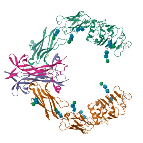
- TrkA (High affinity nerve growth factor receptor) is a receptor tyrosine kinase that serves as the primary receptor for nerve growth factor (NGF). Upon NGF binding, TrkA undergoes dimerization and autophosphorylation, initiating signaling pathways that control neuronal survival, differentiation, and proliferation during nervous system development. Related references: PMID:1281417, PMID:15488758, PMID:17196528
- Downstream signaling cascades include GRB2-Ras-MAPK (cell differentiation and survival), PI3K-AKT (survival), and PLCG1-NF-kappa-B (transcription of survival genes).
- TrkA localizes to the cell membrane and is internalized into early, late, and recycling endosomes upon activation.
- Multiple isoforms exist: TrkA-I is widely expressed in non-neuronal tissues, TrkA-II is neuronal-specific, and TrkA-III is expressed in neural stem/progenitor cells. TrkA-III promotes angiogenesis and has oncogenic activity when overexpressed.
- TrkA is a proto-oncogene subject to autophosphorylation and ubiquitination. It features immunoglobulin-like domains, leucine-rich repeats, and a tyrosine kinase domain.
Experimental Guidance and Technical Tips
- The immunogen (aa 700-796) resides in the intracellular region of TrkA, which may aid in detecting the C-terminal portion of the receptor. Consider this epitope location when planning experimental protocols.
- For Western blotting, denaturing and reducing conditions are typically suitable. Validate with positive controls such as PC12 (rat) or SH-SY5Y (human) cell lysates, observing a band around 87 kDa.
- In immunohistochemistry (IHC-P), antigen retrieval steps may enhance detection. Test on neuronal tissue sections where TrkA expression is expected.
- The antibody’s cross-reactivity with human, mouse, and rat orthologs allows use in diverse preclinical models; confirm specificity in your model system.
- For ELISA applications, consider using recombinant TrkA proteins or cell lysates for assay optimization.
CamelBio: Your One-Stop Sourcing Bridge
CamelBio provides diagnostic manufacturers and research labs with one-stop access to a wide range of IVD raw materials, including validated antibody pairs and optimized polyclonal antibodies like this TrkA Rabbit pAb for neurobiology and oncology target detection. Our portfolio covers bulk ancillary reagents and rare-target sourcing, complemented by technical consulting to accelerate your assay development from concept to clinic.
REQUEST A QUOTE
Our professional team will reply to you within one business day. Please feel free to contact us!