Monoclonal Antibodies
Rabbit anti-Rat CD4 Monoclonal Antibody for Flow Cytometry - P05540
Item Number : CM0007779
Price varies based on specs and customizations
- Application
- FC
- Cross Reactivity
- Rat
- Protein Weight
- 51 kDa

Shipping:
Contact us to get shipping details Enjoy On-time Dispatch Guarantee.
Why Choose Us
Easy ordering process, quality products, and dedicated support for your business success.
Core Product Specifications and Parameters
| Parameter | Value |
|---|---|
| Product Name | Rabbit anti-Rat CD4 mAb |
| Remarks/Alias | Ly49; Klra1; Ly49s2 |
| Species | Rat |
| Gene ID (Human) | 920 |
| Gene ID | 24933 |
| Immunogen | Recombinant fusion protein containing a sequence corresponding to amino acids 1-394 of rat CD4 (NP_036837.1). |
| Source | Rabbit |
| Category | Monoclonal Antibodies |
| Application | FC |
| Cross Reactivity | Rat |
| SWISS | P05540 |
| Protein Weight | 51 kDa |
| Shipping | Ice bag |
Biological Background: CD4 Function and Localization
- CD4, also known as T4/Leu-3 or W3/25 antigen, is a T-cell surface glycoprotein encoded by the Cd4 gene in rat.
- It functions as an integral membrane coreceptor for MHC class II molecules, interacting with the T-cell receptor (TCR) and presenting cells to initiate immune responses.
- Upon MHC engagement, CD4 recruits the Src kinase LCK, leading to phosphorylation cascades that promote T-helper cell activation, lymphokine production, and adhesion.
- CD4 is critical for thymic T-helper cell differentiation and can also trigger monocyte-to-macrophage maturation in an LCK-independent manner.
- Beyond T cells, CD4 expression in macrophages and NK cells influences cytokine expression and cell migration.
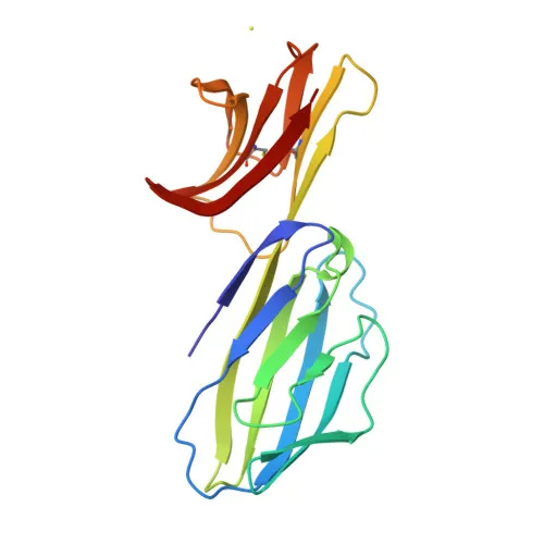
- The protein is located at the cell membrane and features four immunoglobulin-like domains, with a predicted molecular weight of approximately 51 kDa.
- Post-translational modifications include disulfide bond formation, glycosylation, and palmitoylation, which are essential for its structure and function.
- CD4 is a key marker for immune cell subset identification and a primary receptor for HIV entry in humans (structural insights available via 3D-structure keyword).
Experimental Guidance and Technical Tips
- For flow cytometry, titrate the antibody to determine the optimal working concentration for your specific cell population and experimental setup.
- Use appropriate isotype control antibodies conjugated to the same fluorophore () to assess non-specific binding.
- Include a viability dye to exclude dead cells, which can cause false positives due to increased autofluorescence.
- When staining, incubate in the dark at 4°C to minimize antigen modulation and fluorophore photobleaching.
- Since the immunogen comprises amino acids 1-394, the antibody likely recognizes an extracellular epitope, suitable for surface staining without permeabilization. Verify performance if permeabilization is required.
- Validate antibody specificity using CD4-positive (e.g., rat splenocytes or thymocytes) and CD4-negative controls.
CamelBio: Your One-Stop Sourcing Bridge
As a specialized IVD raw material partner, CamelBio supports diagnostic developers and researchers with reliable access to antibodies and reagents essential for immunology and T-cell biology. This Anti-CD4 antibody is part of our extensive portfolio of validated monoclonal antibodies, complemented by bulk ancillary reagents and custom sourcing solutions. From early concept to clinical application, CamelBio streamlines your procurement with one-stop access to critical raw materials.
REQUEST A QUOTE
Our professional team will reply to you within one business day. Please feel free to contact us!