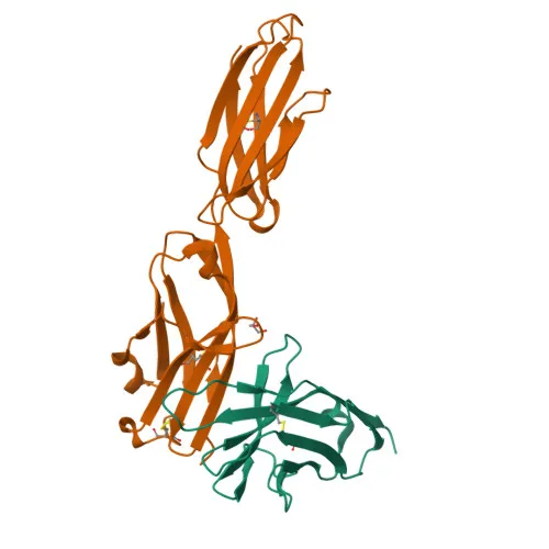
Monoclonal Antibodies
Rabbit anti-Mouse PD-L2/CD273/B7-DC Monoclonal Antibody for Flow Cytometry - Q9WUL5
Item Number : CM0004794
Price varies based on specs and customizations
- Application
- FC
- Cross Reactivity
- Mouse
- Protein Weight
- 28kDa

Shipping:
Contact us to get shipping details Enjoy On-time Dispatch Guarantee.
Why Choose Us
Easy ordering process, quality products, and dedicated support for your business success.
Core Product Specifications and Parameters
| Parameter | Value |
|---|---|
| Product Name | Rabbit anti-Mouse PD-L2/CD273/B7-DC mAb |
| Remarks/Alias | Btdc; B7-DC; PD-L2; F730015O22Rik |
| Species | Mouse |
| Gene ID (Human) | 80380 |
| Gene ID | 58205 |
| Immunogen | Recombinant fusion protein containing a sequence corresponding to amino acids 20-123 of mouse PD-L2/CD273/B7-DC (NP_067371.1) |
| Source | Rabbit |
| Category | Monoclonal Antibodies |
| Application | FC |
| Cross Reactivity | Mouse |
| SWISS | Q9WUL5 |
| Protein Weight | 28kDa |
| Shipping | Ice bag |
Biological Background: PD-L2/CD273/B7-DC Function and Localization
- PD-L2, also known as B7-DC, butyrophilin B7-DC, or CD273, is encoded by the Pdcd1lg2 gene and belongs to the butyrophilin family of immune regulators.
- It plays a critical role in induction and maintenance of immune tolerance to self.
Related references: PMID:11224527, PMID:11283156, PMID:12719480 - Acts as a ligand for the inhibitory receptor PDCD1/PD-1, inhibiting T-cell proliferation by blocking cell cycle progression and cytokine production.
Related references: PMID:11224527, PMID:11283156, PMID:12719480 - PD-L2 is a transmembrane protein localized at the cell membrane.
- It is highly expressed in placenta and liver, with lower expression in heart, spleen, lymph nodes, and thymus; also found on dendritic cells and some tumor cell lines.
- Structural features include an immunoglobulin domain, a transmembrane helix, disulfide bonds, and glycosylation sites.
Experimental Guidance and Technical Tips
- The immunogen covers the extracellular domain (amino acids 20-123), which may help generate antibodies recognizing the native surface protein in flow cytometry.
- For flow cytometry, titrate the antibody to determine the optimal staining concentration and include proper isotype controls.
- Ensure a single-cell suspension and block Fc receptors before staining to minimize non-specific binding.
- The conjugate is compatible with 633-647 nm laser lines; avoid fixatives that may affect fluorophore brightness.
CamelBio: Your One-Stop Sourcing Bridge
CamelBio offers integrated access to high-quality IVD raw materials, expert technical consultation, and end-to-end assay development support. Whether advancing immune checkpoint research or developing diagnostic kits targeting PD-L2, our global supply network can source monoclonal antibodies, validated antibody pairs, and bulk ancillary reagents to meet your project needs. Partner with CamelBio to efficiently bridge the gap from concept to clinical application.
Product Datasheet
Rabbit anti-Mouse PD-L2/CD273/B7-DC Monoclonal Antibody for Flow Cytometry - Q9WUL5
REQUEST A QUOTE
Our professional team will reply to you within one business day. Please feel free to contact us!