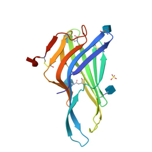
Monoclonal Antibodies
Rabbit anti-Mouse LAMP1/CD107a Monoclonal Antibody for IF/ICC, FC - P11438
Item Number : CM0006197
Price varies based on specs and customizations
- Application
- IF/ICC, FC
- Cross Reactivity
- Mouse
- Protein Weight
- 43kDa

Shipping:
Contact us to get shipping details Enjoy On-time Dispatch Guarantee.
Why Choose Us
Easy ordering process, quality products, and dedicated support for your business success.
Core Product Specifications and Parameters
| Parameter | Value |
|---|---|
| Product Name | Rabbit anti-Mouse LAMP1/CD107a mAb |
| Remarks/Alias | P2B; Perk; LGP-A; CD107a; Lamp-1; LGP-120 |
| Species | Mouse |
| Gene ID (Human) | 3916 |
| Gene ID | 16783 |
| Immunogen | Recombinant fusion protein containing a sequence corresponding to amino acids 208-363 of mouse LAMP1/CD107a (NP_034814.2). |
| Source | Rabbit |
| Category | Monoclonal Antibodies |
| Application | IF/ICC, FC |
| Cross Reactivity | Mouse |
| SWISS | P11438 |
| Protein Weight | 43kDa |
| Shipping | Ice bag |
Biological Background: LAMP1/CD107a Function and Localization
- Lysosome-associated membrane glycoprotein 1 (LAMP1/CD107a) is a highly glycosylated transmembrane protein, also known as LGP-120, P2B, or CD107a.
- Essential for lysosome biogenesis, pH regulation, autophagy, and cholesterol homeostasis, acting as a direct inhibitor of the TMEM175 proton channel to maintain acidic lumen. Related references: PMID:15121881
- In natural killer (NK) cells, LAMP1 participates in cytotoxic granule movement to the cell surface and perforin trafficking to lytic granules, enabling target cell killing.
- Protects NK cells from degranulation-associated damage by preventing self-inflicted injury from their own cytotoxic granule contents. Related references: PMID:23847195
- Presents carbohydrate ligands to selectins and has been implicated in tumor cell metastasis. Related references: PMID:2676155
- Localizes primarily to lysosome and late endosome membranes but also traffics to the cell membrane and cytolytic granule membrane upon activation.
- Post-translational modifications include extensive N-linked glycosylation and disulfide bond formation, critical for its structural stability and function within the lysosomal environment.
Experimental Guidance and Technical Tips
- For immunofluorescence (IF/ICC), use appropriate fixation (e.g., 4% PFA) and permeabilization (e.g., 0.1% Triton X-100) to allow antibody access to intracellular lysosomal compartments. The conjugated fluorophore eliminates the need for a secondary antibody, simplifying the protocol.
- In flow cytometry (FC), LAMP1 is predominantly intracellular under resting conditions but can be detected on the cell surface after cellular activation (e.g., NK cell degranulation). Consider including a cell activation step and using a viability dye to exclude dead cells.
- Validate the antibody in a relevant mouse cell line or primary cell system known to express LAMP1 (e.g., RAW 264.7 macrophages or primary NK cells) and include appropriate isotype controls for FC.
- The immunogen covers amino acids 208–363 of the luminal domain, which may be sensitive to denaturation; avoid over-fixation that could mask the epitope.
CamelBio: Your One-Stop Sourcing Bridge
CamelBio provides diagnostic manufacturers and research laboratories with streamlined access to high-quality IVD raw materials, including validated monoclonal antibodies, bulk ancillary reagents, and rare-target sourcing solutions. This -conjugated anti-LAMP1 antibody supports investigations into lysosome biology, autophagy, NK cell cytotoxicity, and cancer metastasis. Leverage CamelBio’s integrated supply network to accelerate your assay development from concept to clinic.
Product Datasheet
Rabbit anti-Mouse LAMP1/CD107a Monoclonal Antibody for IF/ICC, FC - P11438
REQUEST A QUOTE
Our professional team will reply to you within one business day. Please feel free to contact us!