Monoclonal Antibodies
Rabbit anti-Mouse CD98 Monoclonal Antibody for Flow Cytometry - P10852
Item Number : CM0008158
Price varies based on specs and customizations
- Application
- Flow Cytometry (FC)
- Cross Reactivity
- Mouse
- Protein Weight
- 58kDa

Shipping:
Contact us to get shipping details Enjoy On-time Dispatch Guarantee.
Why Choose Us
Easy ordering process, quality products, and dedicated support for your business success.
Core Product Specifications and Parameters
| Parameter | Value |
|---|---|
| Product Name | Rabbit anti-Mouse CD98 mAb |
| Remarks/Alias | 4F2; Cd98; Ly10; Mdu1; 4F2HC; Ly-10; NACAE; Ly-m10; Mgp-2hc |
| Species | Mouse |
| GeneID (Human) | 6520 |
| GeneID (Mouse) | 17254 |
| Immunogen | Recombinant protein corresponding to amino acids 105-526 of mouse CD98 (NP_032603.3) |
| Source | Rabbit |
| Category | Monoclonal Antibodies |
| Application | FC (Flow Cytometry) |
| Cross Reactivity | Mouse |
| SWISS | P10852 |
| Protein Weight | 58kDa |
| Shipping | Ice bag |
Biological Background: CD98 (SLC3A2) Function and Localization
-
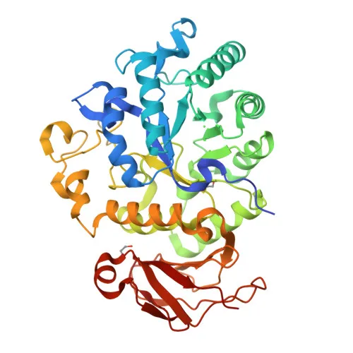
CD98 heavy chain (SLC3A2, 4F2hc) is a type II transmembrane glycoprotein that acts as a molecular chaperone, facilitating the biogenesis, plasma membrane trafficking, and stability of heterodimeric amino acid transporters [PMID:10574970, PMID:10734121, PMID:11011012]. Related references: PMID:10574970 PMID:10734121 PMID:11011012
-
It forms covalent heterodimers via a disulfide bond with various SLC7 light chains, creating functional amino acid exchangers with distinct substrate specificities: system L (large neutral amino acids), system xc− (cystine/glutamate), and system y+L (dibasic amino acids) [PMID:10574970, PMID:10734121, PMID:11011012]. Related references: PMID:10574970 PMID:10734121 PMID:11011012
-
Resides primarily at the plasma membrane (both apical and basolateral domains), but also localizes to lysosome membranes, melanosomes, and cell junctions, consistent with its roles in transport and cell adhesion.
-
Undergoes extensive N‑glycosylation and phosphorylation; contains conserved disulfide bonds critical for its structure and interaction with light chains.
-
Widely expressed in mouse tissues, with highest levels in kidney, small intestine, spleen, thymus, and liver; also detected on brain capillary endothelial cells and embryonic epithelia [PMID:9915839]. Related references: PMID:9915839
-
Beyond transport, CD98 modulates integrin‑dependent signaling pathways, promoting cell spreading, migration, and tumor progression (by similarity).
Experimental Guidance and Technical Tips
- For flow cytometry, use standard surface staining protocols with live cells; no permeabilization required as CD98 is a cell surface antigen.
- Validate antibody performance in the relevant mouse cell type or tissue, taking into account its broad expression; use appropriate isotype controls and Fc block to minimize background.
- As a directly conjugated antibody, minimize light exposure and use fresh aliquots to maintain fluorescence intensity.
- Consider co‑staining with cell type‑specific markers to distinguish CD98‑positive populations.
- Due to potential internalization of CD98, incubations should be at 4°C or on ice to prevent antibody uptake.
CamelBio: Your One-Stop Sourcing Bridge
CamelBio empowers diagnostic developers with one-stop access to high-quality IVD raw materials, including ready-to-use conjugated antibodies like this anti‑CD98 () for immunology and transporter research. Our portfolio spans validated monoclonal/polyclonal antibodies, matched antibody pairs, and ancillary reagents, backed by technical expertise to accelerate your assay development from concept to clinic.
Product Datasheet
Rabbit anti-Mouse CD98 Monoclonal Antibody for Flow Cytometry - P10852
REQUEST A QUOTE
Our professional team will reply to you within one business day. Please feel free to contact us!