Monoclonal Antibodies
Rabbit anti-Mouse CD80 Monoclonal Antibody for Flow Cytometry - Q00609
Item Number : CM0009896
Price varies based on specs and customizations
- Application
- FC
- Cross Reactivity
- Mouse
- Protein Weight
- 34kDa

Shipping:
Contact us to get shipping details Enjoy On-time Dispatch Guarantee.
Why Choose Us
Easy ordering process, quality products, and dedicated support for your business success.
Core Product Specifications and Parameters
| Parameter | Value |
|---|---|
| Product Name | Rabbit anti-Mouse CD80 mAb |
| Remarks/Alias | B71; Ly53; TSA1; Cd28l; Ly-53; MIC17 |
| Species | Mouse |
| GeneID (Human) | 941 |
| GeneID | 12519 |
| Immunogen | Recombinant protein|Recombinant fusion protein containing a sequence corresponding to amino acids 38-245 of mouse CD80(NP_033985.3). |
| Source | Rabbit |
| Category | Monoclonal Antibodies |
| Application | FC |
| Cross Reactivity | Mouse |
| SWISS | Q00609 |
| Protein Weight | 34kDa |
| Shipping | Ice bag |
Biological Background: CD80 Function and Localization
- T-lymphocyte activation antigen CD80 (also known as B7-1) is a costimulatory molecule that belongs to the immunoglobulin superfamily and plays an essential role in T-lymphocyte activation. Related references: PMID:16339518, PMID:1714935
- As a primary auxiliary signal, CD80 augments the MHC/TCR signal in naive T-cells by binding to its receptor CD28, leading to activation of downstream pathways including NF-kappa-B and MAPK.
- CD80 also functions as an inhibitor of T-cell activation by serving as a ligand for CTLA4, thereby blocking CD28-mediated T-cell priming.
- CD28/CD80 costimulation stimulates glucose metabolism and ATP synthesis in T-cells via the PI3K/Akt signaling pathway.
- CD80 acts as a regulator of PDL1/PDCD1 interactions to limit excess engagement of PDL1 and its inhibitory role in immune responses.
- Expressed on B-cells, CD80 is crucial for B-cell/T-cell interactions in both early and late germinal center responses, which are essential for generating effective humoral immune responses. Related references: PMID:22450810
- Subcellular location: Predominantly localized on the cell membrane with single-pass type I membrane protein topology.
- Tissue specificity: Expressed on activated B-cells, gamma interferon-stimulated monocytes, and non-circulating B-cell malignancies.
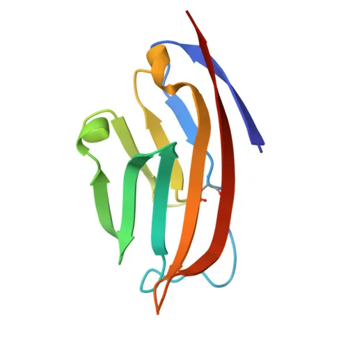
- Post-translational modifications and structural features: Contains a disulfide bond, N-glycosylation, and an immunoglobulin domain; undergoes alternative splicing. Keywords include 3D-structure, glycoprotein, transmembrane helix.
Experimental Guidance and Technical Tips
- The immunogen corresponds to amino acids 38-245, encompassing the extracellular domain of mouse CD80, suggesting the antibody can recognize the native conformation on live cells, ideal for flow cytometry.
- For flow cytometry, consider titrating the antibody to determine optimal concentration, and include appropriate isotype controls and Fc receptor blocking steps.
- As the antibody is pre-conjugated with , it can be used directly for staining without the need for secondary detection reagents.
- Ensure proper handling: store the antibody at recommended temperature (shipped on ice bag) and protect from light to maintain fluorophore stability.
CamelBio: Your One-Stop Sourcing Bridge
At CamelBio, we understand the critical demands of diagnostic development and research supply. As your one-stop sourcing bridge, we offer validated antibody pairs, optimized monoclonal and polyclonal antibodies, and bulk ancillary reagents. For immuno-oncology targets like CD80, CamelBio ensures efficient access to rare raw materials, supporting your journey from concept to clinic.
Product Datasheet
Rabbit anti-Mouse CD80 Monoclonal Antibody for Flow Cytometry - Q00609
REQUEST A QUOTE
Our professional team will reply to you within one business day. Please feel free to contact us!