Monoclonal Antibodies
Rabbit anti-Mouse CD317/BST2 Monoclonal Antibody for Flow Cytometry - Q8R2Q8
Item Number : CM0004805
Price varies based on specs and customizations
- Application
- Flow Cytometry (FC)
- Cross Reactivity
- Mouse
- Protein Weight
- 19 kDa

Shipping:
Contact us to get shipping details Enjoy On-time Dispatch Guarantee.
Why Choose Us
Easy ordering process, quality products, and dedicated support for your business success.
Core Product Specifications and Parameters
| Parameter | Value |
|---|---|
| Product Name | Rabbit anti-Mouse CD317/BST2 mAb |
| Remarks/Alias | GREG; Bst-2; CD317; DAMP-1; 2310015I10Rik |
| Species | Mouse |
| Gene ID (Human) | 684 |
| Gene ID | 69550 |
| Immunogen | Recombinant fusion protein containing a sequence corresponding to amino acids 55-152 of mouse CD317/BST2 (NP_932763.1) |
| Source | Rabbit |
| Category | Monoclonal Antibodies |
| Application | Flow Cytometry (FC) |
| Cross Reactivity | Mouse |
| SWISS | Q8R2Q8 |
| Protein Weight | 19 kDa |
| Shipping | Ice bag |
Biological Background: CD317/BST2 Function and Localization
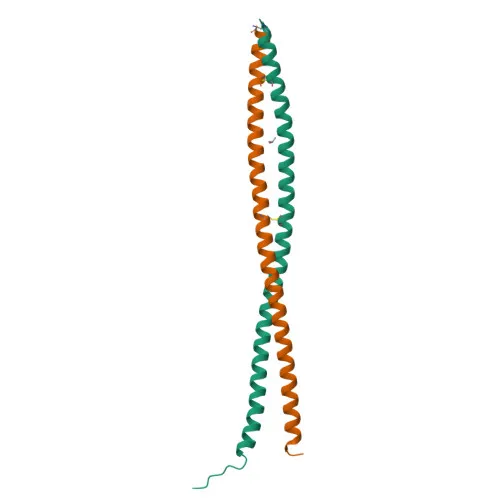
- CD317, also known as bone marrow stromal antigen 2 (BST2), HM1.24 antigen, or tetherin, is an IFN-induced antiviral restriction factor encoded by the Bst2 gene in mouse.
- BST2 directly blocks the release of diverse enveloped viruses from infected cells by tethering nascent virions to the cell membrane via its unique topology—a transmembrane domain and a GPI anchor.
- Targeted viruses include retroviruses (HIV-1, MLV, MMTV), filoviruses (Ebola virus), arenaviruses (Lassa virus), and rhabdoviruses (VSV); tethered virions may be internalized and degraded.
- BST2 can inhibit cell surface proteolytic activity of MMP14, leading to reduced MMP15 activation and subsequently suppressing cell growth and migration.
- BST2 stimulates signaling through LILRA4/ILT7 on plasmacytoid dendritic cells, providing negative feedback to type I IFN production in response to viral infection.
- It plays a role in organizing the subapical actin cytoskeleton in polarized epithelial cells.
- Subcellularly, BST2 localizes to the trans-Golgi network, cell membrane (including membrane rafts and apical membrane), late endosomes, and cytoplasm.
- In naïve mice, BST2 is specifically expressed at the protein level on type I interferon-producing cells, particularly plasmacytoid dendritic cells.
Experimental Guidance and Technical Tips
- The immunogen encompasses the extracellular domain (aa 55-152) of mouse BST2, making this -conjugated antibody suitable for surface staining in live-cell flow cytometry applications.
- For flow cytometry, titrate the antibody to determine the optimal working dilution for your specific cell type and always include appropriate isotype control and fluorescence-minus-one (FMO) controls.
- BST2 expression is known to be induced by type I IFNs; thus, consider stimulating cells with IFN-α/β to enhance signal when detecting endogenous BST2 in certain cell types.
CamelBio: Your One-Stop Sourcing Bridge
CamelBio provides diagnostic manufacturers and research laboratories with streamlined access to high-quality IVD raw materials, including validated antibody pairs, recombinant proteins, and conjugated monoclonal antibodies like this anti-CD317/BST2 clone. With expertise in innate immunity and antiviral restriction factor targets, we offer tailored supply solutions for flow cytometry and immunoassay development. As your one-stop partner, we support every stage from early R&D to scalable clinical reagent delivery, reducing sourcing complexity and accelerating your project timelines.
Product Datasheet
Rabbit anti-Mouse CD317/BST2 Monoclonal Antibody for Flow Cytometry - Q8R2Q8
REQUEST A QUOTE
Our professional team will reply to you within one business day. Please feel free to contact us!