Monoclonal Antibodies
Rabbit anti-Mouse CD317/BST2 Monoclonal Antibody for Flow Cytometry - Q8R2Q8
Item Number : CM0010536
Price varies based on specs and customizations
- Application
- FC
- Cross Reactivity
- Mouse
- Protein Weight
- 19kDa

Shipping:
Contact us to get shipping details Enjoy On-time Dispatch Guarantee.
Why Choose Us
Easy ordering process, quality products, and dedicated support for your business success.
Core Product Specifications and Parameters
| Parameter | Value |
|---|---|
| Product Name | Rabbit anti-Mouse CD317/BST2 mAb |
| Remarks/Alias | GREG; Bst-2; CD317; DAMP-1; 2310015I10Rik |
| Species | Mouse |
| GeneID (Human) | 684 |
| GeneID | 69550 |
| Immunogen | Recombinant fusion protein containing a sequence corresponding to amino acids 55-152 of mouse CD317/BST2 (NP_932763.1) |
| Source | Rabbit |
| Category | Monoclonal Antibodies |
| Application | FC |
| Cross Reactivity | Mouse |
| SWISS | Q8R2Q8 |
| Protein Weight | 19kDa |
| Shipping | Ice bag |
Biological Background: CD317/BST2 Function and Localization
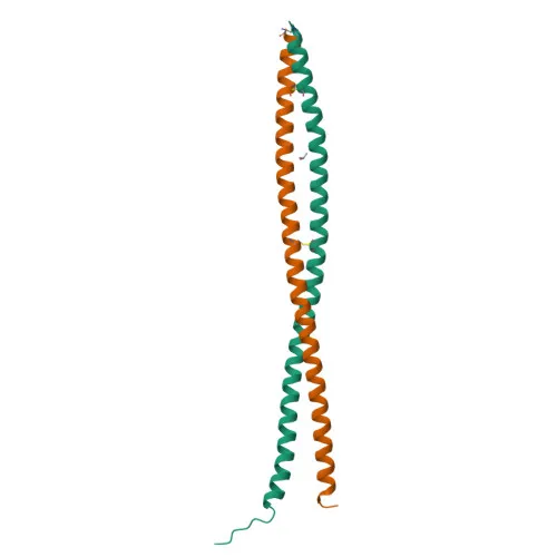
- Bone marrow stromal antigen 2 (BST2), also known as CD317 or HM1.24 antigen, is a 19kDa lipid raft-associated protein with a unique topology including an N-terminal transmembrane domain and a C-terminal GPI anchor.
- Functions as an interferon-induced antiviral restriction factor that blocks release of diverse enveloped viruses (e.g., HIV-1, Ebola, Lassa, VSV, MMTV, MLV) by tethering nascent virions to the cell membrane.
- Exhibits dual topology (type II transmembrane and GPI-anchor) enabling its localization to lipid rafts and facilitating virion tethering and endocytosis for degradation.
- Subcellular localization includes trans-Golgi network, cell membrane, late endosomes, and cytoplasm; polarized expression at the apical membrane in epithelial cells.
- In naive mice, specifically expressed on type I interferon-producing plasmacytoid dendritic cells (at protein level).
- Negatively regulates type I interferon production by plasmacytoid dendritic cells through LILRA4/ILT7 signaling, providing feedback in viral responses.
- Plays a role in organization of subapical actin cytoskeleton in polarized epithelial cells.
- Contains a coiled-coil domain, disulfide bonds, and is N-glycosylated; these post-translational modifications are essential for its structure and antiviral function.
Experimental Guidance and Technical Tips
- The immunogen corresponds to the extracellular domain of mouse CD317 (amino acids 55-152), suggesting the antibody likely targets a surface-exposed epitope suitable for flow cytometry without permeabilization.
- For flow cytometry (FC) applications, use standard staining protocols with appropriate Fc receptor blocking to minimize non-specific binding.
- Consider testing on known BST2-expressing cell types such as mouse plasmacytoid dendritic cells or stimulated epithelial cells; validate in the desired experimental system.
- While validated for FC only, other applications (e.g., immunofluorescence, IHC) may be explored with appropriate optimization and validation in the relevant sample system.
CamelBio: Your One-Stop Sourcing Bridge
CamelBio supplies integrated IVD raw materials and technical services for diagnostic developers and researchers. This rabbit monoclonal antibody targeting mouse CD317/BST2 supports investigations into antiviral immunity and plasmacytoid dendritic cell biology. Beyond this product, CamelBio offers validated antibody pairs, bulk monoclonal and polyclonal antibodies, ancillary reagents, and customized sourcing for rare targets, providing a seamless bridge from concept to clinic.
Product Datasheet
Rabbit anti-Mouse CD317/BST2 Monoclonal Antibody for Flow Cytometry - Q8R2Q8
REQUEST A QUOTE
Our professional team will reply to you within one business day. Please feel free to contact us!