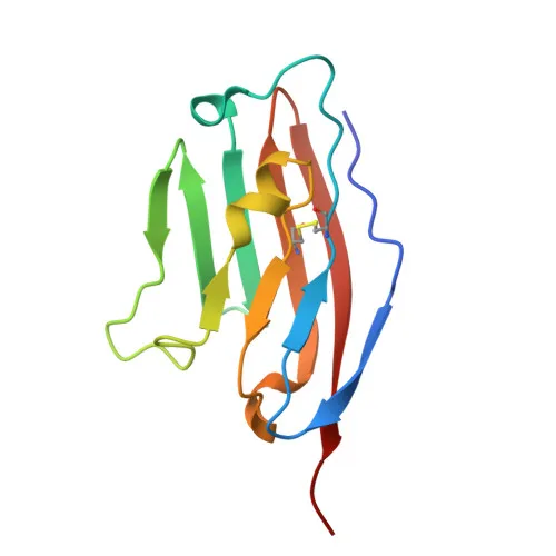
Monoclonal Antibodies
Rabbit anti-Mouse CD274/PD-L1 mAb for Flow Cytometry - Q9EP73
Item Number : CM0012507
Price varies based on specs and customizations
- Application
- FC
- Cross Reactivity
- Mouse
- Protein Weight
- 32kDa

Shipping:
Contact us to get shipping details Enjoy On-time Dispatch Guarantee.
Why Choose Us
Easy ordering process, quality products, and dedicated support for your business success.
Core Product Specifications and Parameters
| Parameter | Value |
|---|---|
| Product Name | Rabbit anti-Mouse CD274/PD-L1 mAb |
| Remarks/Alias | B7h1; Pdl1; Pdcd1l1; Pdcd1lg1; A530045L16Rik |
| Species | Mouse |
| Gene ID (Human) | 29126 |
| Gene ID | 60533 |
| Immunogen | Recombinant fusion protein containing a sequence corresponding to amino acids 19-235 of mouse CD274/PD-L1(NP_068693.1). |
| Source | Rabbit |
| Category | Monoclonal Antibodies |
| Application | FC |
| Cross Reactivity | Mouse |
| SWISS | Q9EP73 |
| Protein Weight | 32kDa |
| Shipping | Ice bag |
Biological Background: CD274/PD-L1 Function and Localization
- Encoded by the Cd274 gene (also known as B7h1, Pdcd1l1, Pdcd1lg1, Pdl1) in mouse, the protein product is formally named Programmed cell death 1 ligand 1, also referred to as B7 homolog 1 (B7-H1).
- Functions as a critical immune checkpoint molecule: interacts with the inhibitory receptor PDCD1/PD-1 to maintain peripheral tolerance by modulating T-cell activation thresholds and limiting effector responses. Related references: PMID:11238124
- Through an alternative, yet unidentified, activating receptor, it may also costimulate specific T-cell subsets that predominantly produce interleukin-10 (IL10). Related references: PMID:11015443 PMID:12719480
- The PDCD1/PD-L1 pathway is frequently exploited by tumors to suppress anti-tumor immunity; ligation inhibits cytotoxic T‑lymphocyte (CTL) effector function, and its blockade can reverse T‑cell exhaustion, underpinning modern cancer immunotherapy strategies. Related references: PMID:12218188 PMID:27281199
- Subcellularly, PD-L1 is primarily anchored at the cell membrane and also cycles through early and recycling endosome membranes.
- Expression is highest in mouse heart, thymus, skeletal muscle, and lung, with lower levels in kidney, spleen, thyroid, and liver; it is also detected on activated dendritic cells, B cells, macrophages, and many lymphoid‑derived tumor cell lines.
- Post‑translational features include glycosylation, a signal peptide, an immunoglobulin V‑like domain, transmembrane helix, disulfide bonds, and ubiquitination sites.
Experimental Guidance and Technical Tips
- This antibody is recommended for flow cytometry (FC) and may detect native surface PD‑L1 on live cells without permeabilization, as the immunogen encompasses the extracellular domain (aa 19–235).
- For FC experiments, consider titrating the antibody (e.g., 0.5–5 µg per test) to determine the optimal staining concentration for your specific cell type and model system.
- Since PD‑L1 expression is inducible and varies with cell activation status, validate detection in both resting and stimulated (e.g., IFN‑γ‑treated) samples.
- Mouse strain and tissue‑specific expression variations should be considered; confirm reactivity in the relevant biological context.
CamelBio: Your One-Stop Sourcing Bridge
CamelBio streamlines IVD raw‑material procurement for diagnostic manufacturers and research laboratories, offering a single‑point access to validated immunoreagents. Our portfolio includes this anti‑mouse PD‑L1 antibody plus complementary antibody pairs, bulk ancillary reagents, and custom sourcing for rare immune‑checkpoint targets—all tailored for robust immunoassay and immunohistochemistry development. Reach out to CamelBio for end‑to‑end support in translating your oncology IVD concepts into reliable clinical tools.
REQUEST A QUOTE
Our professional team will reply to you within one business day. Please feel free to contact us!