Monoclonal Antibodies
Rabbit anti-Mouse CD14 Monoclonal Antibody for FC - P10810
Item Number : CM0010592
Price varies based on specs and customizations
- Application
- FC
- Cross Reactivity
- Mouse
- Protein Weight
- 39 kDa

Shipping:
Contact us to get shipping details Enjoy On-time Dispatch Guarantee.
Why Choose Us
Easy ordering process, quality products, and dedicated support for your business success.
Core Product Specifications and Parameters
| Parameter | Value |
|---|---|
| Product Name | Rabbit anti-Mouse CD14 mAb |
| Remarks/Alias | CD14 |
| Species | Mouse |
| GeneID (Human) | 929 |
| GeneID (Mouse) | 12475 |
| Immunogen | Recombinant fusion protein containing a sequence corresponding to amino acids 18-345 of mouse CD14 (NP_033971.1). |
| Source | Rabbit |
| Category | Monoclonal Antibodies |
| Application | FC |
| Cross Reactivity | Mouse |
| SWISS | P10810 |
| Protein Weight | 39 kDa |
| Shipping | Ice bag |
Biological Background: CD14 Function and Localization
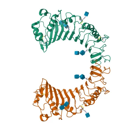
- CD14 is a monocyte differentiation antigen and a myeloid cell-specific leucine-rich glycoprotein, belonging to the leucine-rich repeat (LRR) family and anchored to the membrane via a glycosylphosphatidylinositol (GPI) moiety.
- Acts as a coreceptor for bacterial lipopolysaccharide (LPS), binding monomeric LPS in concert with LBP and delivering it to the LY96/TLR4 complex to initiate innate immune responses. Related references: PMID:16148141
- LPS-CD14 signaling proceeds via the MyD88/TIRAP/TRAF6 pathway, leading to NF-kappa-B activation, cytokine secretion, and inflammatory responses. Related references: PMID:15895089, PMID:8612135
- Also functions as a coreceptor for bacterial lipopeptides: synergizes with TLR2:TLR6 for diacylated lipopeptides and with TLR2:TLR1 for triacylated lipopeptides; signaling occurs from the cell surface, followed by lipid-raft-dependent trafficking to the Golgi.
- Serves as an accessory receptor for M. tuberculosis lipoproteins LprA, LprG, and LpqH, cooperating with TLR2 and TLR1 to modulate antigen-presenting cell functions. Related references: PMID:19362712
- Localizes to the cell membrane, membrane rafts, and Golgi apparatus; a soluble secreted form is also generated.
- Expressed on peritoneal macrophages and lung alveolar/dendritic macrophages at the protein level. Related references: PMID:8612135, PMID:19362712
- Post-translationally modified: contains a GPI-anchor attachment site, disulfide bonds, and N-linked glycosylation, contributing to its structural stability and function.
Experimental Guidance and Technical Tips
- For flow cytometry, titrate the antibody to determine the optimal working concentration for your cell system; typical starting dilutions range from 1:50 to 1:200.
- Use appropriate isotype controls and consider Fc receptor blocking to reduce nonspecific binding, especially when working with myeloid cells.
- If intracellular staining is needed, fix and permeabilize cells after surface staining; note that CD14 is primarily surface-expressed but may internalize upon ligand binding.
- Validate staining on known CD14-positive populations, such as peritoneal macrophages or lung alveolar macrophages, to confirm specificity and sensitivity.
CamelBio: Your One-Stop Sourcing Bridge
CamelBio provides diagnostic manufacturers and research laboratories with integrated access to premium IVD raw materials and technical expertise. This anti-mouse CD14 monoclonal antibody supports innate immunity and inflammation studies, and we can supply validated antibody pairs, optimized bulk reagents, and custom conjugation services. Our one-stop sourcing model helps you secure all necessary raw materials efficiently, accelerating development from concept to clinical application.
REQUEST A QUOTE
Our professional team will reply to you within one business day. Please feel free to contact us!